
Being pregnant often feels like waiting for the best surprise of your life. You wonder what your baby looks like, who they will resemble, and what their little features might be. A 4D ultrasound scan brings you closer to the answer, showing live video of your baby inside the womb.
What is a 4D ultrasound scan?
4D ultrasound creates real-time videos of your baby, rather than just still pictures. While the familiar black-and-white 2D ultrasound shows snapshots, 4D ultrasound captures movements as they happen. The "4D" part refers to time as the fourth dimension, so you can see length, width, depth, and motion all at once.
While doctors primarily rely on 2D ultrasounds for routine monitoring and detecting anomalies such as cleft lip, 4D scans can help enhance visualisation of certain features, like the face or hands.
Essential prenatal tests like blood tests and growth scans provide medical reassurance about your baby's health. A 4D ultrasound, while not medically necessary, can offer an emotional bonding experience for parents.
What is the difference between 2D, 3D and 4D ultrasound scans?
Each type of ultrasound presents a different level of detail and purpose. Here’s how they compare:
2D ultrasound:
The scan most parents are familiar with, showing black-and-white images in slices. Doctors use it to check your baby’s growth, organs, and overall development. It is the primary tool for monitoring health and detecting structural anomalies such as cleft lip or heart defects.
3D ultrasound:
This scan creates still images in three dimensions, providing clearer views of your baby’s features, such as the nose, mouth, hands, and feet. While 2D scans are medically sufficient, 3D can help enhance visuals of certain features and make some conditions easier to see.
4D ultrasound:
Think of this as 3D in motion. Instead of a still picture, you see a real-time video of your baby moving, stretching, or making facial expressions. 4D scans are mainly optional for parents to see their baby in action. Doctors may use them occasionally to assist in visualising features, but routine monitoring and anomaly detection are done with 2D scans.
When is a 4D ultrasound scan performed?
Getting the timing right makes all the difference between clear and amazing footage, and disappointing images. This happens between week 26 and 34 weeks of pregnancy, typically during the second trimester to third trimester.
Why this timing works so well:
Your baby has developed enough fat to look adorable (not like a tiny skeleton)
Facial features are completely formed and clearly visible
There's still enough room to see your baby's whole body moving around
Amniotic fluid levels are usually just right for clear viewing
If you have the scan too early, your baby won’t have enough subcutaneous fats to give a clear picture. If you wait too late, your baby might be too big and too low to see the face well. That’s why doctors help you pick the best time for the scan.
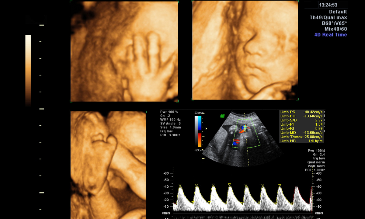
What can you see during the scan?
During the scan, parents can enjoy a moving video of their baby inside the womb. You may see:
Stretching or turning
Thumb sucking
Yawning
Changing facial expressions
The scan also reveals detailed facial features and limbs, giving parents a lifelike first glimpse of their baby while doctors continue to observe healthy movement and bonding moments.
Want to experience the joy of seeing your baby before birth? Request a 4D ultrasound appointment and take home videos or images to treasure these special moments.
What is the role of 4D ultrasound scan during pregnancy?
4D ultrasounds create real-time video of your baby, showing movements and expressions as they happen. While they're not typically part of routine prenatal care, they can serve both medical and emotional purposes when used appropriately.
Medical applications
While 2D ultrasounds remain the standard for most pregnancy monitoring, 4D ultrasounds can be helpful for assessing specific conditions that are harder to see in traditional scans, such as:
Facial development:
Detecting cleft lip or other facial differences more clearly
Foetal behavior assessment:
Observing movement patterns that may indicate neurological development
Complex structural issues:
When 2D images need additional visualization
Bonding and emotional benefits
4D ultrasounds provide parents with a valuable opportunity to view and bond with the unborn baby. Many parents find watching their baby's real-time movements, like yawning, stretching, or moving their hands, creates a deeper emotional connection during pregnancy.
When are they recommended?
Your doctor might suggest a 4D ultrasound if:
There's a specific concern that needs enhanced visualization
Traditional 2D images aren't providing clear enough information
You're at higher risk for certain conditions that benefit from detailed imaging
Our O&G specialists
Loading...
What to expect during the appointment?
Before your ultrasound examination, doctors may give simple advice:
Stay hydrated to improve image quality
Eat or drink something cold or sweet about 20 minutes before may encourage your baby to move
Wear loose clothing for comfort
Bring your partner to share the moment
During the scan:
You’ll lie back on the couch, and warm gel will be spread over your tummy
The sonographer will move a small probe across your skin
On the screen, you’ll see your baby in real time like movements, facial features, even little expressions
The sonographer may also take a few measurements of your baby’s head, tummy, and limbs
At the end, the gel is wiped away, and you’ll be given copies of the images or video
Is 4D ultrasound safe?
A 4D ultrasound scan is considered safe. It uses harmless sound waves, not radiation. This is the same principle as the obstetric ultrasound used for routine prenatal care. When performed by trained professionals, there is no evidence of risk to mother or baby.
Limitations of 4D ultrasound scan
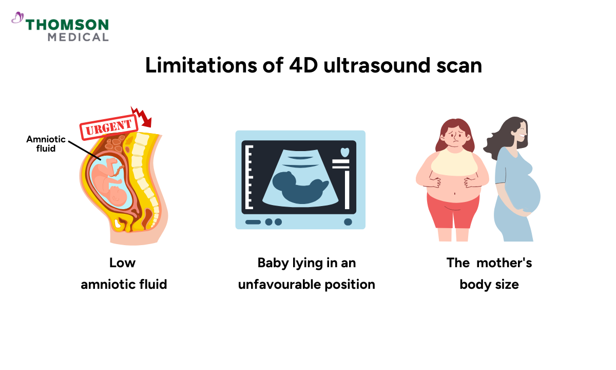
Although exciting, a 4D ultrasound has some limits. Image quality can be reduced by:
Low amniotic fluid
Baby lying in an unfavourable position
The mother's body size
If these factors are present, the scan may not provide clear views.
FAQ
Is a 4D ultrasound scan necessary during pregnancy?
No, it is optional. Routine care relies on 2D ultrasound scans, especially the mid-pregnancy anomaly scan. A 4D ultrasound is usually chosen for bonding and extra reassurance. It is often combined with other prenatal tests to give a fuller picture of your baby’s health.
How is 4D ultrasound different from a regular scan?
A regular ultrasound examination is typically 2D. It checks structures, growth, and fetal anomalies. A 4D ultrasound scan adds live video, showing movements and expressions in real time.
When is the best time to get a 4D fetal ultrasound?
The ideal time is 26 to 34 weeks. At this stage, your baby has enough fat under the skin for recognisable facial features, but is still small enough for clear views.
Will a 4D ultrasound scan detect abnormalities?
Yes, it can reveal some major concerns, such as issues affecting the heart, skull, limbs, or abdomen. However, the standard second-trimester anomaly scan remains the most important test for detecting fetal abnormalities.
What affects the quality of a 4D ultrasound image?
Clarity depends on a few factors. Low amniotic fluid levels, your baby’s position, or the mother’s body size can all make it harder to see clear images.
What happens if the images aren’t clear?
If this happens, the sonographer may ask you to move, or adjust the probe. Sometimes drinking more water helps. If your baby stays in an awkward position, the appointment might be rescheduled.
The information provided is intended for general guidance only and should not be considered medical advice. For personalised recommendations and tailored advice based on your unique situations, please consult a specialist at Thomson Medical. Request an appointment with Thomson Medical today.
For more information, contact us:
Thomson Specialists (Women's Health)
Thomson Women's Clinic (TWC)
- Novena:
6592 6686 (Call), 8611 8986 (WA) - Bukit Batok:
6569 0668 (Call), 8686 3525 (WA) - Choa Chu Kang:
6893 1227 (Call), 8282 1796 (WA) Jurong:
6262 8588 (Call), 6262 8588 (WA)- Katong (female doctor):
6970 2272 (Call), 8611 9020 (WA) - Punggol:
6243 6843 (Call), 8811 0328 (WA) - Sembawang: 6753 5228
- Sengkang: 6388 8125
- Serangoon (female doctor): 6382 3313
- Tampines: 6857 6266
- Tiong Bahru: 6276 1525