
Have you ever wondered how your doctor can see inside your heart? Doctors use a cardiac MRI scanner, which is a specialised version of magnetic resonance imaging. This scan provides a detailed view of your heart using powerful magnets and radio waves, making the procedure painless and free from radiation.
What is a cardiac MRI?
A cardiac MRI scan is a non-invasive medical imaging technique that creates detailed, high-quality images of your heart and blood vessels. This imaging can assess the structure of your heart (such as the chambers, valves, and muscles) and evaluate how well they function. The scan can also assess how well blood flows through your blood vessels.
Since powerful magnetic fields and radio waves are used to create the images, there is no risk of radiation exposure, unlike with X-rays or CT scans. This means that this cardiac imaging is safe for repeated assessments when necessary. Cardiac MRI scans also offer better soft tissue detail than other medical imaging methods.
These scans can provide two- or three-dimensional images, helping your healthcare provider to diagnose, monitor, and guide treatment for various cardiovascular conditions. When greater clarity is required, your doctor may use a contrast agent called gadolinium to highlight specific structures or blood flow patterns.
When does the doctor recommend a cardiac MRI?
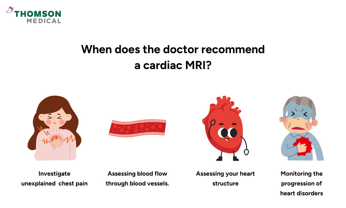
Your doctor may recommend a cardiac MRI scan if they suspect an issue with your heart health and need to evaluate its structure and function. Your doctor may recommend this cardiac imaging in the following situations:
Investigate unexplained heart conditions such as chest pain, shortness of breath, or fainting
Assessing blood flow through the heart and major blood vessels, including monitoring for pulmonary hypertension (elevated blood pressure in the lungs)
Monitoring the progression of heart disorders over time
Evaluating heart valve disorders such as regurgitation (heart valve leakage) or stenosis (heart valve narrowing)
Assessing your heart structure, such as its size, muscle thickness, and the function of your heart chamber
Diagnose conditions such as coronary artery disease, myocarditis (heart inflammation), or cardiomyopathy (heart muscle disease)
Assess congenital heart defects in children or adults born with heart abnormalities
Diagnosing the cause of heart failure or any unexplained changes in the heart function
Detecting any irritation or inflammation of the pericardium (the outer membrane covering your heart)
Assessing for any damage to your heart or areas that lack blood flow due to heart artery blockages caused by a heart attack (myocardial infarction)
Furthermore, this medical imaging is used to formulate treatment plans for your heart conditions and assess the effectiveness of the treatment.
If you are experiencing symptoms such as chest pain, are at risk of heart disease, or require monitoring of an existing cardiac condition, a cardiac MRI scan can help uncover the underlying cause. Schedule an appointment with Thomson Medical for a thorough evaluation and a personalised treatment plan.
How do I prepare for a cardiac MRI?
Preparing for an MRI scan is relatively straightforward, and your healthcare provider will give you instructions to help you prepare. A radiology technologist, a professional with training and certification to operate the MRI equipment, performs this imaging test.
Here are some general guidelines to help you prepare:
Clothing:
Before the scan, you may be asked to change into a hospital gown and remove any jewellery, dentures, watches, or other metal objects that might interfere with it.
Metal objects:
Inform the technologist if you have any metal implants, such as pacemakers, stainless steel intrauterine devices, or other medical devices, as they can heat up and pose a safety risk to you.
Medications:
Bring a list of any medications you are taking, as some may need to be temporarily stopped before the procedure.
Avoid caffeine:
It's recommended that you skip any caffeinated beverages, such as coffee, tea, or energy drinks, for at least 24 hours before the scan. This is because caffeine can affect your heart rate, potentially leading to inaccurate results.
Contrast dye:
If your doctor orders a contrast-enhanced MRI, you may need to be injected with a contrast dye (gadolinium) to improve the visibility of your heart and blood vessels.
Pregnancy:
If you are pregnant or think you might be, tell your doctor or technologist before the scan.
If you are anxious about being in enclosed spaces (claustrophobia), talk to your healthcare provider. You may be given a sedative before the procedure to help you relax during the scan
Additionally, if you have a kidney condition and your scan requires contrast dye, inform your healthcare provider. They may take a blood test before the scan to check your kidney function and assess whether it is safe to use a contrast dye.
Please don't hesitate to ask your doctor or technologist any questions you may have. They will be delighted to answer your questions and ensure that your cardiac MRI goes smoothly and safely, giving you peace of mind.
How does the test work?
Following the preparation stage above, the procedure will begin and usually lasts 30 to 90 minutes. During the scan, you can expect the following:
If a gadolinium contrast agent is required before the scan, your technologist will inject it through an IV into your arm vein.
You will then be asked to lie down on a table that slides into a large, tubular machine that looks like a tunnel.
Then a small sticky patch with an electrocardiogram (ECG) lead will be placed on your chest to monitor your heartbeat during the scan.
When the scan starts, the MRI machine will make loud tapping or thumping sounds. You'll be given earplugs or headphones to wear before the procedure to protect your hearing.
It's important to stay still during the scan to ensure clear images. You may be asked to hold your breath for a short time.
If there are issues, you can use the intercom to contact the MRI radiologist in another room.
If you didn't have a sedative for the MRI scan, you will not need a recovery period. You can return home and resume your normal activities. However, if you have had a sedative for the scan, you will need to recover from its effects before you can go home safely.
If your scan requires a contrast dye, you will usually need to drink plenty of fluids to help flush the dye through your urine.
What to expect after the procedure?
A radiologist will analyse the images and send a signed report to your doctor. Your doctor will then share the results with you and discuss everything with you.
If a follow-up examination is required, your doctor will explain why, which may include further evaluation using different imaging methods or monitoring changes over time. Follow-up scans are often recommended to assess the effectiveness of treatment or to monitor any changes in your condition.
If you would like more information about cardiac MRI scans, schedule a consultation with Thomson Medical. Our specialists can explain this procedure to you in more detail and answer any questions you might have.
What are the potential risks of this medical imaging test?
Although MRI scans are generally safe, there are some potential risks you need to be aware of:
The loud noise produced by the MRI scanner can be uncomfortable, particularly if the scan takes more than an hour
If you're someone who gets anxious in small spaces (claustrophobia), you might feel uneasy during your MRI scan.
If your scan requires a contrast agent, some people may rarely experience an allergic reaction to gadolinium. Symptoms may include nausea, a metallic taste in the mouth, and an allergic reaction such as rashes, flushed skin, and shortness of breath.
People with metal implants, pacemakers, or other devices may need special precautions or can't have an MRI.
You may feel nervous or stressed while lying still in the MRI machine for a long periods of time
FAQ
What does an MRI of your heart show?
A cardiovascular imaging provides detailed, three-dimensional images of your heart's structure and shows how it functions. It enables doctors to assess:
- Heart muscle health:
- Detects damage from heart attacks, inflammation (myocarditis) or scarring
- Heart size and shape:
- It reveals whether the heart is enlarged or if the walls are abnormally thick or thin
- Heart valves:
- Checks whether the valves are functioning properly or if they are leaking (regurgitation) or narrowed (stenosis)
- Blood flow:
- Identifies issues with blood flow, such as blockages, weak spots or abnormalities in arteries and veins
- Congenital heart disease:
- Helps diagnose structural heart problems present from birth
Which is better, an MRI or a CT scan for the heart?
Cardiac MRI and cardiac CT scans serve different purposes. Cardiac CT is ideal for detecting calcium buildup (plaque) in the arteries and identifying coronary artery disease (blockages) and provides fast results, often within minutes.
Cardiac MRI, on the other hand, provides a more detailed view of the heart muscle, valves and blood flow. It is particularly useful for diagnosing heart inflammation, scarring, and congenital heart defects, as well as for evaluating heart function after a heart attack.
In general, a CT scan is preferred for quickly detecting artery blockages, while an MRI scan is used for a comprehensive assessment of the heart muscle and its function.
Can MRI detect heart blockage?
A cardiac MRI scan can reveal blood flow problems in the heart, which may indicate a blockage. However, it is not typically the first test used to diagnose coronary artery disease. If a blockage is suspected, your doctor may recommend a coronary CT scan or a stress test first.
Is a cardiac MRI better than an echocardiogram?
Both of these cardiovascular imaging methods have their strengths, and the most suitable one depends on what the doctor is looking for.
- Echocardiogram (echo):
- It uses ultrasound, is quick and non-invasive, and is excellent for assessing heart valve function, heart motion, and overall pumping ability.
- Cardiac MRI:
- An MRI scan offers significantly more detailed images of the heart muscle, valves, and blood flow, enabling the detection of scar tissue, inflammation, and complex heart conditions.
- An MRI scan is especially helpful when an echocardiogram does not provide enough detail or clarity.
If a quick check-up is needed, an echocardiogram is ideal. However, if doctors need a detailed, in-depth look at the heart, an MRI scan is better.
Who should get a cardiac MRI?
Your doctor may recommend a cardiac MRI for the following reasons:
- Suspected heart damage from a past heart attack
- Unexplained chest pain that needs further investigation
- Heart muscle diseases like cardiomyopathy
- Heart inflammation (myocarditis) or scarring
- Congenital heart defects (problems with heart structure from birth)
- Valve disease that needs closer examination
What are the side effects of a heart MRI?
A cardiac MRI scan is generally safe and does not involve radiation. Most people experience no side effects. However, some people may occasionally experience mild discomfort from lying still or an allergic reaction to gadolinium contrast agent (if used), such as a cool sensation or mild itchiness.
If you have kidney problems, your doctor will carefully consider whether it is appropriate for you to have contrast dye. Serious reactions are rare.
The information provided is intended for general guidance only and should not be considered medical advice. For personalised recommendations based on your medical conditions, schedule a consultation with Thomson Medical.
For more information, contact us:
Thomson Medical Concierge
- 8.30am - 5.30pm
- WhatsApp: 9147 2051
Need help finding the right specialist or booking for a group?
Our Medical Concierge is here to help you. Simply fill in our form, and we'll check and connect you with the right specialist promptly.
Notice:
The range of services may vary between Thomson clinic locations. Please contact your preferred branch directly to enquire about the current availability.
Get In Touch