
If your doctor has mentioned getting an MRI or fMRI scan, you might feel confused about what these tests involve. Both scans help doctors see inside your body without surgery, but they look for different things. Knowing the differences can help you feel more prepared and less anxious about your upcoming scan.
What is MRI?
Magnetic Resonance Imaging (MRI) is a safe, non-invasive imaging technique that produces highly detailed pictures of the inside of your body. Unlike X-rays or CT scans, the MRI does not use ionising radiation, which makes it particularly useful for repeated imaging.
MRI is widely used to help diagnose and monitor a broad range of conditions affecting various parts of your body. For instance:
Brain and spinal cord:
MRI can detect brain strokes, injuries blood vessel damage, tumours (including cancer), multiple sclerosis, and spinal cord injuries.
Heart and blood vessels:
It helps identify heart disease, blocked blood vessels, and structural abnormalities in your heart.
Bones and joints:
MRI is effective in diagnosing joint disorders, disc problems in the spine, bone infections, and certain cancers.
Other organs:
The technique is also used to examine the liver, kidneys, breasts, pancreas, pelvis, and prostate for abnormalities or disease.
How does an MRI work?
An MRI scan works by using a strong magnetic field and radio waves to create images of your body’s internal structures. Here’s how it works:
Magnetic field alignment:
First, the MRI machine generates a strong magnetic field.
This magnetic field causes the hydrogen atoms in your body’s water and fat molecules to align with the MRI's magnetic field due to their magnetic properties.
Radio wave pulse:
The MRI machine then sends a radio frequency (RF) pulse into your body.
This pulse temporarily knocks the hydrogen atoms out of their new alignment.
Signal emission:
When the RF pulse is turned off, these hydrogen atoms gradually return to their original alignment. As they realign, they emit faint radio signals.
Signal detection and image creation:
Sensors (coils) inside the MRI machine detect these emitted signals. A computer then processes the signals from different parts of the body.
By analysing the timing and strength of the signals, the computer constructs detailed cross-sectional images (slices) of your body’s internal tissues.
What is fMRI?
Functional magnetic resonance imaging (fMRI) is a safe, non-invasive imaging method that measures and maps brain activity. It works by detecting changes in blood flow and oxygen levels within the brain, which increase when specific regions become active during thinking, movement, or sensory processing.
It helps your doctors identify which areas of your brain are involved in different tasks, map out brain connections, and study changes in brain activity due to conditions such as stroke, brain injury, or neurological diseases.
In clinical practice, functional MRI scans are especially valuable before brain surgery, as they allow doctors to pinpoint active or critical brain regions, helping to plan safer and more effective procedures. Additionally, fMRI assists in understanding the impact of strokes or other disorders on the brain or spinal cord.
How does an fMRI work?
An fMRI works by detecting changes in blood flow that occur when brain activity increases. Functional MRIs record evidence of brain behaviour based on changes in neural activity that occur in one or more parts of the brain during all human activities.
The process relies on a simple but important fact about how your brain works. Here's how the fMRI captures your brain activity:
The most common fMRI method is called Blood Oxygen Level Dependent, or BOLD. When you perform any physical or intellectual task, the active areas of your brain receive more blood and oxygen. The fMRI scanner then detects these increases in blood and oxygen levels, which indirectly measure neural activity.
Areas of the brain involved in a particular activity will show evidence of greater blood flow by generating stronger-than-usual signals during an fMRI scan. Brighter areas on fMRI scan results indicate brain activity in regions associated with movement or remembering. Darker areas indicate parts of the brain that are not in use or in a resting state.
BOLD is a relative method, comparing oxygen levels during active periods with those during rest. This comparison allows doctors to see exactly which brain areas become more active when you perform different tasks.
To find out more about fMRI and MRI imaging, schedule a consultation with Thomson Medical. Our specialist can provide you more information about this procedure and decide which scan is best for you.
fMRI vs. MRI: What are the differences?
Both imaging techniques use magnetic fields and radio waves to create detailed images, but they serve different purposes
| Feature | MRI | fMRI |
|---|---|---|
| Primary purpose | Obtain detailed images of your body's anatomy, including bones, soft tissues, organs, and muscles | Measure brain activity by detecting changes in blood flow level in your brain |
| What it shows | It focuses on structural information, which means it provides detailed images that show how your body parts look, including bones, organs, and soft tissues. | It focuses on functional information, which means it provides insights into how your brain works by mapping and measuring activity in different brain regions during tasks or at rest. |
| Body areas | It can scan any part of your body, such as brain, spine, joints, organs, muscles | Exclusively focuses on your brain and brain activity |
| Type of images | Creates still "snapshot" images at a single moment in time | Creates dynamic images showing brain activity over time |
| What you do during scan | You lie still and quiet throughout the entire scan | Although you’ll also be required to lie still, you’ll be asked to perform tasks like thinking, moving fingers, or answering questions while being scanned |
| What it detects | Physical abnormalities, such as injuries, tumours, structural damage, inflammation | Brain function, such as which areas are active, blood flow changes, functional problems |
| Common uses | • Diagnosing joint injuries • Detecting tumors • Checking organ health • Examining spinal problems • Looking for structural brain damage | • Planning brain surgery • Studying epilepsy • Research on brain functions • Mapping brain activity • Understanding brain disorders |
What is it like to have an MRI or fMRI test? Comparing MRIs and fMRIs
Knowing what to expect during your scan can help you feel more prepared and less anxious.
Your MRI experience
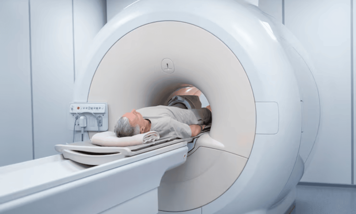
During your MRI, you'll lie on a padded table that slides into a large, tube-shaped machine. The procedure is completely painless, you won't feel anything happening inside your body during the medical imaging scan.
During the scan, the MRI machine will produce loud, rhythmic sounds. These sounds are normal and just mean the machine is working properly. Your technician will provide you earplugs or headphones to make the noise more tolerable and protect your hearing.
You'll be asked to lie still throughout the scan so the pictures come out clear and sharp. Some people may feel claustrophobic inside the MRI machine due to the enclosed space, but remember that you can communicate with your technician at any time if you need help.
Your fMRI experience
The experience of having an fMRI is similar to an MRI in many ways. You'll lie on the same type of padded table that slides into the same tube-shaped machine. The machine noise is the same as an MRI, those same loud, rhythmic sounds, and you'll receive the same ear protection.
However, there are some important differences during your fMRI. You may be asked to engage in specific tasks, such as thinking about certain topics or moving parts of your body, to activate brain areas during the scan. These tasks might include:
Moving your fingers or toes when instructed
Thinking about specific memories or images
Looking at pictures on a screen
Listening to sounds or words
Answering simple questions
Although you are engaged in these activities, it is essential to remain as still as possible during the scan. The tasks are designed to be simple and manageable while lying in the machine. These activities help the scanner see which parts of your brain become active during different types of thinking and movement.
What are the disadvantages or risks?
While MRI and fMRI scans are generally safe procedures, it's important to know the potential risks and limitations. Most people experience no problems during their scan, but being informed helps you prepare and feel more comfortable.
MRI risks
Claustrophobia:
The enclosed space of the MRI scanner may cause discomfort or anxiety for some individuals, especially those with claustrophobia.
Loud noise:
MRI machines produce loud tapping or knocking sounds during the scan, which can be unpleasant. However, earplugs or headphones are typically provided to reduce discomfort.
Metal implants or devices:
While not a direct health risk, certain metal implants (like pacemakers, cochlear implants, or some aneurysm clips) may be incompatible with MRI due to the strong magnetic field.
In such cases, your healthcare provider will assess whether an MRI is safe for you or recommend an alternative imaging method.
fMRI risks
Same as MRI:
Functional MRI (fMRI) involves the same scanning process and therefore carries the same risks as a standard MRI, including claustrophobia, sensitivity to noise, and incompatibility with some metal implants.
Limitations in brain activity measurement:
fMRI does not measure brain activity directly. Instead, it detects changes in blood flow, which may not perfectly represent actual neuronal activity.
Task performance challenges:
fMRI often requires you to perform specific physical or mental tasks during the scan.
For individuals with cognitive impairments or difficulty following instructions, completing these tasks may be challenging, affecting the accuracy of the results.
FAQ
What does an fMRI offer that an MRI does not?
While MRI gives us a clear picture of your brain's anatomy, fMRI reveals:
Which areas of your brain become active during specific tasks
How different brain regions communicate with each other
How your brain responds to various stimuli or activities
Patterns of brain activity that may indicate certain conditions
Regular MRI shows us the structure, but fMRI shows us the brain at work.
What is an fMRI used for?
Functional magnetic resonance imaging (fMRI) is primarily used to observe and analyse brain activity, revealing which areas of the brain are involved in various tasks or thought processes. It is commonly applied in:
Neuroscience research:
To explore cognitive functions and understand how the brain works during different mental activities.
Clinical assessment:
To evaluate brain activity in patients with conditions such as epilepsy, stroke, or brain injuries.
Pre-surgical planning:
To help surgeons identify and avoid critical brain regions before performing operations.
Mental health studies:
To gain insight into mental processes and investigate disorders like anxiety or depression.
Can fMRI detect brain damage?
fMRI can identify areas where brain function has been affected by damage from conditions like:
Stroke
Traumatic brain injury
Neurological disorders
However, fMRI primarily shows how well different brain areas are functioning rather than revealing physical damage. For the clearest picture of brain health, doctors often use both MRI and fMRI together. The MRI shows any structural damage, while the fMRI reveals how that damage affects brain function.
Is fMRI better than MRI?
Neither fMRI nor MRI is universally "better"—each serves a different purpose. MRI provides highly detailed images of the body’s anatomy, including the brain, organs, and bones, making it ideal for diagnosing structural abnormalities.
fMRI, on the other hand, is specialised for mapping brain activity by detecting differences in blood flow, so it is better suited to understanding how the brain works during specific tasks or at rest. The choice depends on what information your doctor needs.
Who cannot use fMRI?
MRI and fMRI scans are not recommended for people with certain implanted devices, such as pacemakers, cochlear implants, or neurostimulators, unless the device is specifically labelled as MRI-safe or MRI-conditional. Patients with metal fragments, clips, or other metal objects in their bodies should also avoid MRI unless cleared by their healthcare professional.
Can an fMRI detect all types of brain injury?
fMRI is highly sensitive to changes in brain activity and can detect certain types of functional abnormalities, such as those caused by concussions or strokes, that might not be visible on a standard MRI. However, some structural injuries or subtle damage may still be better seen with a traditional MRI.
The information provided is intended for general guidance only and should not be considered medical advice. For personalised recommendations based on your medical conditions, schedule a consultation with Thomson Medical.
For more information, contact us:
Thomson Medical Concierge
- 8.30am - 5.30pm
- WhatsApp: 9147 2051
Need help finding the right specialist or booking for a group?
Our Medical Concierge is here to help you. Simply fill in our form, and we'll check and connect you with the right specialist promptly.
Notice:
The range of services may vary between Thomson clinic locations. Please contact your preferred branch directly to enquire about the current availability.
Get In Touch