
What is a foot MRI?
A foot MRI is a non-invasive imaging test that provides your healthcare provider with detailed images of the soft tissues in your foot and ankle. During the scan, an MRI machine utilises radio waves and strong magnetic fields to capture images of your foot and ankle structures.
This imaging technique allows your doctor to identify a wide range of conditions, including fractures, dislocations, sprains, tendon ruptures, cartilage injuries, and other potential foot and ankle problems.
When is a foot MRI used?
Your healthcare provider may suggest a foot MRI if you're experiencing symptoms in your foot or ankle that need a closer look. These symptoms might include:
Pain that won't go away
Swelling in your foot or ankle
Redness or warmth in the area
Numbness or tingling sensations
Stiffness or reduced sensation in your foot
You cannot put weight on your foot without pain.
Your doctor will typically recommend an MRI when your X-ray imaging don't show any clear injury, but you're still having problems that need to be investigated further.
What can a foot MRI detect?
An MRI scan can see things that X-rays might miss. This detailed scan can help your doctor identify several different conditions:
Bone problems
Bone bruising or bone contusion
Small cracks in your bones (stress fractures)
Partial breaks that don't show up on X-rays
Synovial based disorders
Inflammation of the synovial membrane (synovitis - which causes joint swelling)
Inflamed tendons (tenosynovitis - inflammation of connective tissues that protects your tendons)
Swollen fluid sacs near your joints (bursitis)
Small fluid-filled lumps (ganglion cysts)
Other Conditions
Congenital or developmental abnormalities (dysplasia)
Infections in your bones, joints, or soft tissues (osteomyelitis, osteoarthritis)
Unusual growths or tumours in your bones, joints, or soft tissues
Plantar fasciitis, which happens when the thick band of tissue on the bottom of your foot becomes irritated and swollen.
Achilles tendinitis, which happens when the tendon that connects your calf muscle to the heel bone becomes irritated or inflamed.
The MRI gives your doctor a detailed picture of what's happening inside your foot, helping them create the right treatment plan for you.
For more information about foot MRI and to determine if it’s appropriate for your condition, consider speaking with a healthcare professional. You may contact Thomson Medical to arrange a consultation for personalised guidance tailored to your individual care needs.
How do I prepare for a foot MRI?
The best way to prepare for your foot MRI is to discuss the details with your healthcare provider beforehand. Different testing facilities may have slightly different preparation requirements, so it's important to get specific instructions for where you'll be having your test done.
Your healthcare provider will give you a complete physical examination before your MRI. This knowledge helps ensure the test is safe and appropriate for you. You'll need to tell your doctor about several important things:
Any medical conditions you currently have
All medications you're taking (including prescription and over-the-counter drugs)
Any supplements or vitamins you take regularly
Any allergies you have to medications
Any medical implants you have (such as pacemakers, artificial heart valves, artificial joints, cochlear implants, or surgical screws)
Previous surgeries where metal objects may have been used
If you are pregnant or think you might be, please note that pregnant women may need special considerations before imaging.
On the day of your test, wear loose, comfortable clothing that doesn't have:
Zippers
Buttons
Metal snaps or fasteners
This helps avoid any interference with the MRI machine and makes the process more comfortable for you.
What happens before, during, and after a foot MRI scan?
Before the scan, you’ll be asked to remove all metal items, such as jewellery, keys, and watches, because metal can interfere with the MRI machine. These items must be left outside the scan room. You may also be asked to change into a hospital gown or wear loose-fitting clothing without any metal parts.
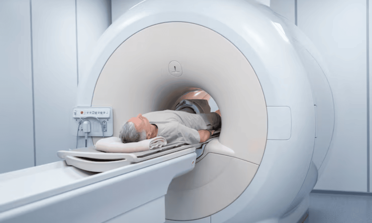
During the scan, you’ll lie down on the MRI table, usually with your feet pointing into the machine. Your foot will be positioned carefully to ensure accurate imaging. It’s important to lie still during the scan, as movement can affect the scan image quality.
The MRI machine can be quite loud while it’s running, so you may be offered earplugs or headphones. In some cases, music may be played to help you relax. You might feel slight warmth in your foot during the scan—this is normal and not harmful.
An intercom system allows you to speak with the radiographer at any time, so you won’t be left alone or unheard during the procedure.
If no additional tests are required after the scan, you can proceed to dress and return home. After reviewing the results, your healthcare provider will discuss them with you.
How does the test work?
An MRI scan is a safe, non-invasive imaging test that doesn't require any cuts or needles to see inside your body. Instead, it uses radio waves and a strong magnetic field to take detailed pictures of your foot and ankle.
While X-rays are great for showing bones, an MRI gives your doctor much more detailed images of your soft tissue structures. These soft tissues include:
Muscles
Tendons (which connect muscles to bones)
Ligaments (which connect bones to other bones)
Cartilage (the smooth tissue that cushions your joints)
Blood vessels
This detailed view of your soft tissues is especially important when you have foot or ankle problems. Many injuries and conditions affect these soft tissues rather than just the foot bones, so the MRI helps your doctor see exactly what's causing your symptoms.
The comprehensive pictures from your MRI scan give your healthcare provider the information they need to make an accurate diagnosis and create the best treatment plan for you.
What are the potential risks and side effects of this test?
Your foot MRI scan is generally very safe. Most people have no problems at all during or after the test. However, it's helpful to know about some potential issues you might experience so you can be prepared.
Loud noise:
The MRI machine makes loud knocking, tapping, or buzzing sounds while it's taking pictures. This is completely normal, but it can be startling if you're not expecting it. You'll be given earplugs or headphones to help reduce the noise.
Allergic reaction:
If your test requires contrast dye (a special liquid that helps create clearer images), you might experience:
Mild nausea (feeling sick to your stomach)
Dizziness
Hives (small, itchy bumps on your skin)
Flushed or warm feeling in your skin
These reactions are usually mild and go away quickly. Serious allergic reactions are very rare.
Claustrophobia:
Some people feel uncomfortable in the MRI machine because it's an enclosed space. This feeling is called claustrophobia. If you think this might bother you, let your healthcare team know beforehand – they can help make you more comfortable.
Stress or anxiety:
The process can cause emotional discomfort for some, especially if you're nervous about medical tests.
FAQ
What does an MRI of the foot show?
Your foot MRI creates detailed, high-resolution images of all the internal structures inside your foot and ankle. This includes your muscles, tendons, ligaments, nerves, and other tissues that X-rays can't see clearly.
These detailed images help your doctor spot injuries, inflammation, or other problems that might be causing your symptoms.
How long does a foot MRI usually take?
Most ankle scans or foot MRIs take around 30 to 60 minutes, depending on the number of views your doctor needs and whether a contrast dye is required. These scan times may vary slightly, and your radiology team will let you know what to expect before the entire scan begins.
Do you have to go all the way in for a foot MRI?
No, you don't! For your foot MRI, only your feet and lower legs need to go inside the machine. Your head and upper body will stay outside the scanner, which makes the experience much more comfortable. This is different from other types of MRI scans where you might go in headfirst.
Can MRI show nerve damage in the foot?
Yes, your MRI can show if there are problems affecting the nerves in your foot. It can detect swelling, compression, or other structural issues that might be pinching or damaging your nerves. This scan helps your doctor understand if nerve problems are contributing to your pain, numbness, or tingling.
Which is better, an MRI or a CT scan for a foot?
That depends on what your doctor is looking for. An MRI provides detailed imaging of soft tissues, making it ideal for diagnosing common conditions like ligament sprains, ankle tendon injuries, or inflammation. However a CT scan is better at showing bone abnormalities, bone fractures, cracks in bones, and detailed views of tarsal bones and other bones in joints.
Since many foot problems involve soft tissue injuries, your doctor will often recommend an MRI first. Your healthcare provider will explain why they're choosing one test over the other based on your symptoms.
What should I expect after my foot MRI is complete?
Once your ankle scans are complete, you can immediately resume your normal activities. Your scanning image will be reviewed by a radiologist who specialises in reading detailed, high-resolution images of internal structures.
They'll create a report about any common conditions, foot or ankle bone injuries, or ankle conditions they find, which will be sent to your doctor.
Your healthcare provider will then contact you to discuss the results and, if needed, develop an effective treatment plan to improve your foot health and ankle health. The entire process from scan to results typically takes a few days to a week.
The information provided is intended for general guidance only and should not be considered medical advice. For personalised recommendations based on your medical conditions, request an appointment with Thomson Medical.
For more information, contact us:
Thomson Medical Concierge
- 8.30am - 5.30pm
- WhatsApp: 9147 2051
Need help finding the right specialist or booking for a group?
Our Medical Concierge is here to help you. Simply fill in our form, and we'll check and connect you with the right specialist promptly.
Notice:
The range of services may vary between Thomson clinic locations. Please contact your preferred branch directly to enquire about the current availability.
Get In Touch