
What is a meniscus?
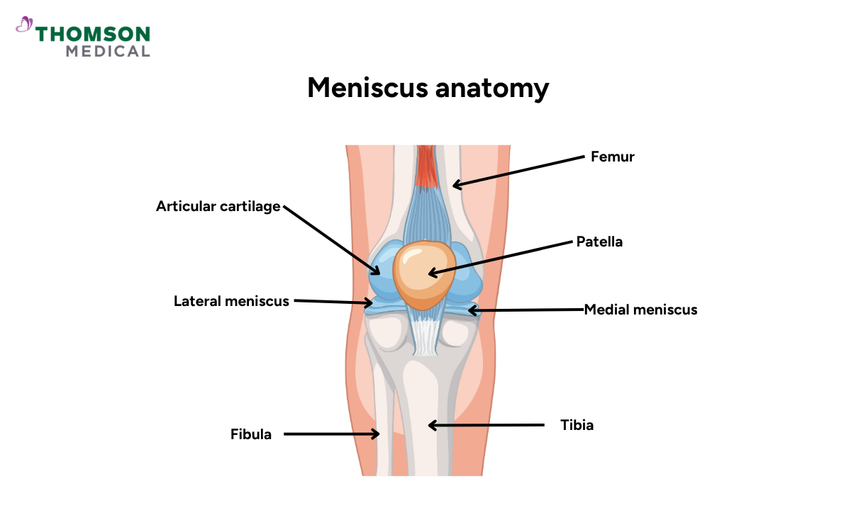
The knee meniscus is two C-shaped pieces of cartilage that sit between the thigh bone (femur) and the shin bone (tibia) in each knee. There are two menisci in each knee, the medial meniscus on the inside and the lateral meniscus on the outside.
Medial vs lateral meniscus
Medial meniscus:
Located on the inner side of the knee, it is more C-shaped, almost semicircular and less mobile, making it more prone to injury.
Lateral meniscus:
Located on the outer side, it is tighter and closed off like an incomplete circle.
It is slightly smaller than a medial meniscus and is more mobile, which offers some protection against tears.
Basic structure and function
The knee menisci are:
Concave on the top (facing the femur) and flat on the bottom (resting on the tibia).
Composed mainly of fibrocartilage, which provides both flexibility and strength.
The knee menisci are attached to small grooves in the top of the tibia, called fossae, located between the areas where the femur rests (tibial condyles). They thin out like a narrow shelf in the middle of the knee joint, unattached to the bone.
The meniscus facilitates stability and ensures that weight is evenly distributed in the knee joint. The primary role of the meniscus is to act as a ”cushion” or shock absorber during high-impact activities, protecting the lower part of the leg.
The meniscus also enables smooth knee movement by providing lubrication to the knee joint and preventing the knee bones from rubbing against each other.
Why understanding meniscus anatomy helps with better treatment outcome
Understanding meniscus anatomy is beneficial for both doctors and patients alike. A detailed understanding of meniscus anatomy enables doctors to:
Accurately diagnose and treat knee injuries
Accurately identify tear locations
Predict the likelihood of healing (based on blood supply)
Tailor different treatment plans for the patients (e.g., conservative treatment or surgical treatment)
Thoroughly plan for meniscus surgery when needed
Interpret different imaging methods, such as an X-ray scan or a Magnetic Resonance Imaging (MRI) scan, more effectively.
A detailed understanding of meniscus anatomy enables you to:
Grasp the potential consequences of meniscus injuries like meniscus tears
Understand the rationale behind the different treatment options
Prevent future injuries from occurring
Zonal anatomy of the meniscus
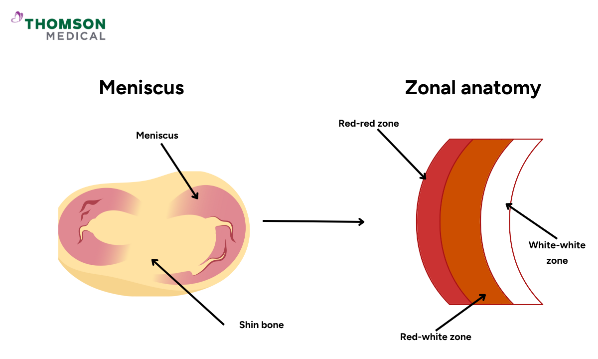
The blood flow in the meniscus travels from the periphery (outside) part to the central meniscus. As people age, blood flow decreases, and by adulthood, the central meniscus becomes lacking in blood supply, which slows down the healing process.
The meniscus is divided into three zones based on blood supply, which directly impacts healing potential:
Red-red zone (outer third-vascular)
Location:
Outer third of the meniscus
Blood supply:
Receives the most blood supply compared to the other zones
Healing potential:
High healing potential after meniscus repair surgery due to the abundance of blood flow, which supports tissue repair
Red-white zone (intermediate vascularity)
Location:
Middle third of the meniscus
Blood supply:
Receives some blood supply, but not as much as red-red zone
Healing potential:
Variable; some capacity for healing after meniscus repair surgery, but less than the red-red zone and dependent on tear size and age of patient
White-white zone (inner third-avascular)
Location:
Inner third of the meniscus, closest to the centre of the knee.
Blood supply:
No blood supply, nourished by the joint fluid in knee
Healing potential:
Poor, as there is no direct blood supply to support repair. Usually treated with a partial meniscectomy since meniscus repair is not possible.
Why vascular zones matter for tear healing
Tears in the red-red or red-white zones are more likely to heal on their own or with surgical repair due to better blood supply.
Tears in the white-white zone rarely heal on their own, as this area relies solely on diffusion from joint fluid for nourishment. Therefore, surgical intervention is necessary.
What are the common types of meniscus tears?
Radial tear:
The tear extends from the inner edge outward, perpendicular to the curve.
Horizontal tear:
Splits the meniscus into upper and lower sections, parallel to the tibial surface.
Vertical (Longitudinal) tear:
The longitudinal tear often follows the curve of the meniscus, typically in the vascular outer third.
Bucket handle tear:
A displaced longitudinal tear where a portion of the meniscus is displaced, forming a handle-like fragment.
Flap tear:
A loose piece of meniscus tissue forms a flap that can catch in the joint.
Complex tear:
Combines features of several tear types, resulting in a fragmented or irregularly shaped tear.
The information provided is intended for general guidance only and should not be considered medical advice. If you have any concerns about a potential meniscus tear, please consult a specialist at Thomson Medical. Request an appointment with Thomson Medical today.
How to spot the meniscus tear location?
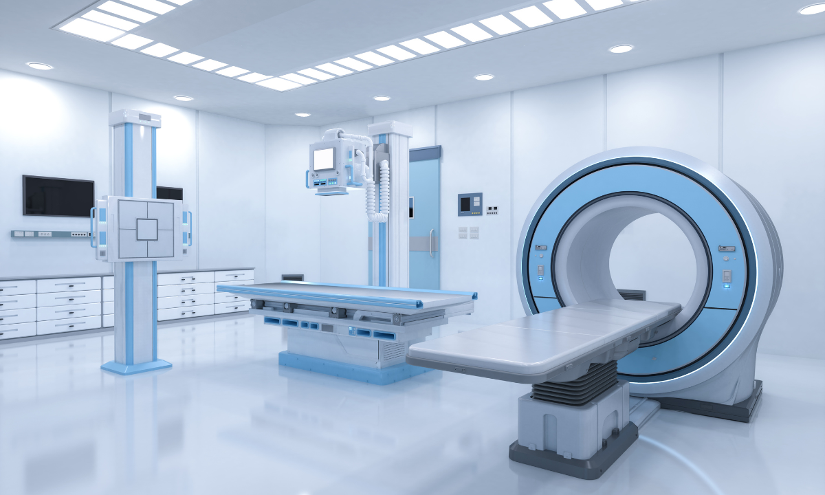
Your doctor will conduct a thorough evaluation of your knee and review your medical history if they suspect a meniscal tear. They may also order X-rays and magnetic resonance imaging (MRI) to confirm the diagnosis and further evaluate the knee joint
X-ray scan:
An X-ray is a diagnostic test that uses invisible electromagnetic energy beams to produce images of internal tissues, bones, and organs onto film.
When a standard X-ray is not accurate enough, a joint X-ray with contrast dye may also be used to examine your knee joints.
X-rays are useful for ruling out other potential knee problems that could cause similar symptoms, such as fractures or arthritis.
MRI scans:
An MRI is a diagnostic procedure that uses a combination of large magnets, radiofrequencies, and a computer to produce detailed images of organs and structures within the body and can often determine damage or disease in a surrounding ligament or muscle.
This will help visualise the tear’s exact location, orientation, and depth, as well as determine which vascular zone is involved.
A torn meniscus will look disrupted, and you may see bright, white vertical or horizontal lines through the meniscus. There may also be some fluid build-up within or around the meniscus.
Since X-rays do not show soft tissues such as cartilage like the meniscus, MRI scans are preferred to diagnose meniscus tears.
How do doctors grade meniscus changes?
Meniscus tears are graded according to their severity:
Grade 0 means that your meniscus is normal without any tear
Grade 1, this grade indicates a mild, small tear, usually in the outer side of the meniscus.This condition is often asymptomatic and may heal naturally.
Grade 2, this level indicates a deeper tear, often located in the red-white zone, where healing occurs more slowly.
Grade 3, refers to acomplete tear of the meniscus, which typically requires surgical intervention.
FAQ
How many menisci are in the knee, and what are their names?
There are two menisci in each knee, the medial meniscus (inner side) and the lateral meniscus (outer side).
Which meniscus is more commonly torn?
The medial meniscus is more frequently injured due to its reduced mobility and stronger attachment to surrounding structures.
What’s the difference between the medial and lateral meniscus?
The medial meniscus is C-shaped and less mobile, while the lateral meniscus is more circular and mobile.
What is the significance of the red-red, red-white, and white-white zones?
These zones indicate the blood supply to different parts of the meniscus, which determines the potential for healing after injury.
Why do some meniscus tears heal on their own while others don’t?
Tears in areas with good blood supply (red-red and red-white zones) can heal naturally, while those in avascular regions (white-white zone) rarely heal without intervention.
How do doctors tell if a meniscus tear is serious or not?
Severity is assessed by symptoms, physical examination, and MRI findings. Tears that are large, unstable, or in avascular zones are considered more serious and less likely to heal without treatment.
Can you tear the same meniscus twice?
Yes, it is possible to re-tear the same meniscus, especially if the initial tear did not heal fully or if the knee sustains further trauma.
The information provided is intended for general guidance only and should not be considered medical advice. For personalised recommendations and tailored advice, please consult a specialist at Thomson Medical. Request an appointment with Thomson Medical today.
For more information, contact us:
Thomson Specialists (Thomson Medical Centre) — Orthopaedic
Request an Appointment