
What is a knee MRI?
A knee MRI scan is a diagnostic imaging test used to examine the internal structures of your knee joint. It provides a comprehensive view of soft tissues, enabling your healthcare provider to assess parts of your knee, such as the ligaments (e.g. ACL, PCL, MCL, LCL), menisci, articular cartilage, muscles, tendons, blood vessels, and nerve pathways.
This level of detail makes MRI the gold standard for diagnosing a wide range of knee injuries and abnormalities, including ligament tears, meniscus injuries, cartilage degeneration, fluid buildup, tumours, and inflammatory conditions.
By offering clear, detailed images, a knee MRI helps clinicians determine the underlying cause of knee pain, swelling, instability, or limited motion and plays a crucial role in planning appropriate treatment plans. Knee MRI scans are available at public hospitals, private hospitals, and imaging centres across Singapore.
How is a knee MRI scan commonly used?
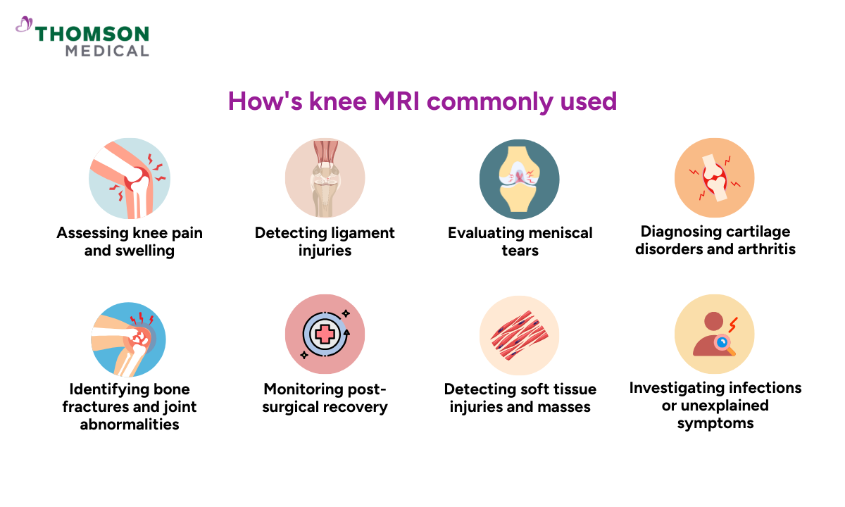
Knee MRI scans are valuable diagnostic tools that allow healthcare providers to visualise soft tissues and bone structures in detail. They are often used to evaluate the cause of knee symptoms and guide treatment decisions. Here are some conditions where a knee MRI can be used, such as:
Assessing knee pain and swelling:
MRI helps identify the underlying causes of persistent pain, inflammation, or swelling that do not show up on X-rays.
Conditions like joint effusion, synovitis, or inflammation can be clearly seen.
Detecting ligament injuries:
An MRI can visualise tears or sprains in major ligaments, including the ACL, PCL, MCL, and LCL.
These injuries are commonly associated with sports-related injuries and sudden movements.
Evaluating meniscal tears:
The menisci (cartilage cushions between the thigh and shin bones) are prone to injury from twisting or overuse.
MRI is the most accurate method to detect meniscus tears, damage or degeneration.
Diagnosing cartilage disorders and arthritis:
MRI reveals thinning, softening, or wear of articular cartilage, which is associated with arthritis and its common types, such as osteoarthritis.
Furthermore, this imaging test can identify early-stage cartilage damage that an X-ray might miss.
Identifying bone fractures and joint abnormalities:
Occult or stress fractures that are not visible on X-rays can be clearly seen with MRI. It can also help identify congenital joint abnormalities or post-traumatic deformities.
Monitoring post-surgical recovery:
MRI is used to evaluate the success of procedures like ACL reconstruction or meniscal repair.
This imaging test is also used to monitor the healing process and to detect complications such as graft failure or the formation of scar tissue.
Detecting soft tissue injuries and masses:
Muscle or tendon strains, cysts like Baker’s cyst, and tumours near the knee joint can be evaluated.
MRI can differentiate between benign and potentially harmful soft tissue changes.
Investigating infections or unexplained symptoms:
MRI may detect joint infections, abscesses, or unexplained knee dysfunction. This is particularly useful in diagnosing cases of fever, redness, or persistent swelling.
Early detection can make a significant difference. Request an appointment with us to find out whether a knee MRI scan is right for you and get a comprehensive check for knee injuries and other knee damages.
When should you have a knee MRI?
A knee MRI is typically recommended when a more detailed evaluation of the internal structures of the knee is needed—especially when initial imaging methods like X-rays or physical examinations do not provide a clear diagnosis. Your healthcare provider may advise a knee MRI in the following situations:
Persistent or unexplained knee pain:
If you experience chronic knee pain that does not improve with rest, medication, or physiotherapy, an MRI can help identify subtle or hidden causes such as cartilage damage or soft tissue injuries.
Acute injury from sports or accidents:
If your doctor suspects a torn ACL, PCL, MCL, or meniscus tear, an MRI provides high-resolution images to confirm the diagnosis and guide treatment.
Swelling or instability:
Unexplained swelling, locking, or a sensation that the knee might “give way” could point to structural damage.
An MRI can help assess whether there's a tear, loose body, or joint effusion causing these symptoms.
Pre- and post-surgical evaluation:
If surgery is being considered, an MRI helps plan the procedure by showing the extent of damage. It’s also useful after surgery to monitor healing or check for complications.
Suspected tumours or infections:
Although less common, an MRI might be necessary to investigate unusual symptoms such as a constant deep ache, fever, or weight loss, which could indicate infection or a bone tumour.
Failure to respond to initial treatments:
When conservative treatments like physiotherapy, medication, or injections don’t lead to improvement, an MRI can uncover hidden issues not seen on X-ray or ultrasound.
Whether it’s a torn ligament, cartilage damage, or unexplained swelling, a detailed scan can uncover the root cause and help guide your next steps toward lasting relief. Request an appointment with us for a thorough evaluation and to determine whether an MRI for knee assessment may be necessary.
What does a damaged knee look like on MRI?
An MRI scan provides detailed images of the internal structures of the knee, making it a powerful tool for identifying various types of knee damage. Depending on the specific injury, the appearance on MRI can vary:
Ligament injuries (e.g., ACL, PCL, MCL, LCL):
Tears may appear as a discontinuity or complete absence of the normal dark band representing the ligament.
Partial tears show as swelling, thickening, or irregular signals within the ligament; complete tears may show retraction (pulling back) of the torn ends and surrounding fluid.
Meniscal tears:
The menisci normally appear as dark, wedge-shaped structures.
A tear appears as an abnormal bright line (high signal) extending to the surface of the meniscus.
Common tear patterns seen include horizontal, radial, complex, or bucket-handle tears.
In some cases, a meniscus tear may also lead to a parameniscal cyst in your knee, which appears on MRI as a small fluid-filled pocket next to the meniscus.
Cartilage damage (articular cartilage):
Thinning or loss of cartilage appears as irregular surfaces or areas of signal loss.
Focal defects may expose the underlying bone (subchondral bone), indicating advanced degeneration or osteochondral injuries.
Bone marrow changes (bone bruising or fractures):
Show as areas of increased brightness (high signal) on fluid-sensitive MRI sequences, indicating bone marrow oedema.
Stress fractures or osteochondral lesions may appear as fine lines or defects in the bone surface.
Joint effusion and swelling:
Excess fluid in the joint space appears bright on MRI scans.
Synovitis (inflammation of the joint lining) may present as thickened synovial tissue, sometimes enhanced further with contrast.
Tendon and muscle injuries:
Tendon problems (like tendinopathy or tendonitis) show up as thick or inflamed areas on the scan.
Tears in tendons look like gaps or breaks in the tissue.
Muscle injuries may show signs of bleeding, swelling, or damaged muscle fibres.
Other findings:
Baker’s cysts: Fluid-filled swellings at the back of the knee, visible as bright, well-defined areas.
Loose bodies: Small fragments of bone or cartilage floating within the joint space.
Chronic injuries: May appear as scar tissue, degenerative changes, subchondral cysts, or calcification.
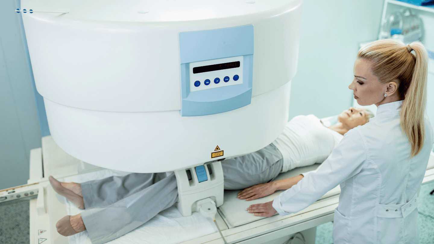
What is the procedure for a knee MRI?
Here’s what you can expect during the procedure:
Before the scan
You’ll be asked to remove any metal items, such as jewellery, keys, or watches.
You may be given a gown to wear for comfort and safety.
If your doctor has ordered a contrast-enhanced MRI, a contrast dye may be injected into a vein in your arm to help highlight certain tissues in the knee.
During the scan
You will lie down on your back on a sliding table.
Your knee will be gently positioned in a cushioned support to keep it stable and in the correct alignment.
The table will then move into the MRI machine, which is a large, tunnel-like scanner.
It’s important to stay as still as possible to ensure clear images of your knee.
The scanner makes loud tapping or knocking sounds, but you’ll be provided with earplugs or headphones to minimise the noise.
The scan usually takes 20 to 30 minutes for a standard knee MRI, or slightly longer if contrast dye is used.
After the scan
Once the MRI test is complete, you can resume normal activities right away.
If contrast dye was used, you’ll be advised to drink water to help flush it out of your system.
A radiologist will review your knee images and send a report to your doctor, who will then explain the results and next steps.
Choosing between an X-ray and an MRI
X-rays: Fast and effective for visualising bones
X-rays are typically the first imaging method used to assess bone-related issues in the knee, such as fractures, dislocations, or joint degeneration like osteoarthritis. They provide quick and clear images of dense structures like bones and joint spaces.
X-rays are widely available, low-cost, and ideal for emergency or initial evaluations. However, they do not show soft tissue injuries such as ligament tears or cartilage damage.
MRI scans: Best for detailed imaging of soft tissues
MRI scans offer superior image clarity for soft tissues in and around the knee. They are ideal for identifying ligament injuries (e.g. ACL, PCL), meniscal tears, cartilage damage, and tendon problems.
MRI is often recommended when knee pain or swelling is unexplained by X-ray findings. As MRI does not use radiation, it is also suitable for long-term monitoring or for younger patients. However, it is more costly and may not be as readily available as X-rays.
Doctors often start with an X-ray for bone injuries or arthritis and move on to an MRI if soft tissue damage is suspected. While X-rays are quicker and more accessible, MRIs provide a more comprehensive view for deeper knee problems. The choice will depend on the nature of your symptoms, your medical history and your doctor's clinical judgement.
Not sure which scan is right for your knee concerns? Request an appointment with us today, and our orthopaedic specialists will guide you through the most suitable imaging option and ensure you get the right diagnosis for effective treatment.
Price of an MRI scan for knee detection in Singapore
The cost of a knee MRI scan in Singapore varies depending on a variety of factors. These include the physician, whether or not contrast dye is used, and whether the scan is conducted in a public or private facility.
At subsidised rates in public hospitals, an MRI scan ranges between SGD 400 and SGD 1000. This may be higher in some instances for non-subsidised patients or referrals from private clinics.
Private facilities or hospitals range from about SGD 900 upwards to more than SGD 2,500 depending on the complexity of the scan, as well as whether specialist consultation is included.
Some important factors that may affect the total cost include:
The need for specialist interpretation or follow-up consultation
Urgency of the scan
Inclusion of pre- or post-scan services
Patients may use MediSave to offset part of the cost, especially if the scan is medically indicated. However, coverage amounts and eligibility vary, so it is advisable to check with your healthcare provider or the relevant institution beforehand.
The information provided above is intended for general reference only. For detailed fee information and payment options, please consult your healthcare provider directly. Request an appointment with our specialists at Thomson Medical today for a detailed price breakdown and a personalised care plan.
FAQ
What does a knee MRI show?
A knee MRI uses strong magnetic fields and radio waves to produce detailed images of the soft tissues inside your knee. This includes structures such as the ligaments, menisci, cartilage, muscles, blood vessels, and nerves.
It is especially useful for detecting knee injuries, such as ligament tears or meniscal damage, as well as identifying other abnormalities like inflammation, degeneration, or structural issues that may not be visible on X-rays.
How long does a knee MRI take?
A knee MRI scan, including preparation and post-scan steps, typically takes about 30 to 60 minutes. However, if contrast dye is used, the scan may take slightly longer.
Do you have to go all the way in for a knee MRI?
No, in most cases, you don’t have to go fully into the MRI machine for a knee MRI. Usually, only your lower body or knee needs to be positioned inside the scanner, while the rest of your body remains outside. The scan is focused specifically on the internal structures of your knee, making it a more comfortable experience for most patients.
Will an MRI show all knee damage?
A knee MRI can detect most types of knee damage, especially involving soft tissues such as ligaments, menisci, cartilage, tendons, and surrounding muscles. However, an MRI may not reveal all issues on its own.
To provide a complete and accurate diagnosis, doctors typically use it in conjunction with a thorough medical history, physical examination, and sometimes additional tests like X-rays. Together, these tools help doctors better understand the root cause of your knee problem and plan the most effective treatment.
What are the disadvantages of a knee MRI?
While a knee MRI is a highly effective and non-invasive imaging tool, it does come with a few potential drawbacks:
Costly compared to other imaging tests
Can cause discomfort for those with claustrophobia
Risk of allergic reaction to contrast dye (if used)
Loud noise during the scan, though ear protection is provided
Will an MRI show an old knee injury?
Yes, an MRI can detect old knee injuries, especially if they resulted in structural changes such as ligament scarring, meniscal tears, cartilage wear, or bone bruising. Even if the injury occurred years ago, an MRI may still reveal signs of chronic damage or degeneration related to the past trauma.
However, the clarity of these signs may vary depending on the severity of the injury and how well it healed.
What does not show up on a knee MRI?
While a knee MRI provides detailed images of soft tissues and bones, there are some things it may not clearly show:
Minor inflammation or early arthritis changes that are more functional than structural
Subtle infections or metabolic conditions without clear tissue damage
Past pain or discomfort that has no visible physical cause
Microscopic damage or very early-stage tissue changes
Certain foreign bodies, especially small fragments of wood or plastic, which may not be visible
Are there any risks associated with a knee MRI scan?
Knee MRI scans are generally safe, but possible risks include:
Loud noise during the scan
Allergic reactions to contrast dye, such as nausea or skin irritation
Claustrophobia in some patients
Mild stress or anxiety during the procedure
Therefore, you should always let your doctor know if you have concerns before the scan.
The information provided is intended for general guidance only and should not be considered medical advice. For personalised recommendations and tailored advice, please consult a specialist at Thomson Medical. Request an appointment with Thomson Medical today.
For more information, contact us:
Thomson Medical Centre MRI Suite
- Mon-Fri: 9am - 5pm
- Sat: 9am - 1pm
Contact Our Patient Care Team
- 8.30am - 5.30pm
- WhatsApp: 9147 2051
- We provide diagnostic MRI examinations performed by qualified radiographers.
- All MRI examinations require a doctor's referral. Fees are available upon request.