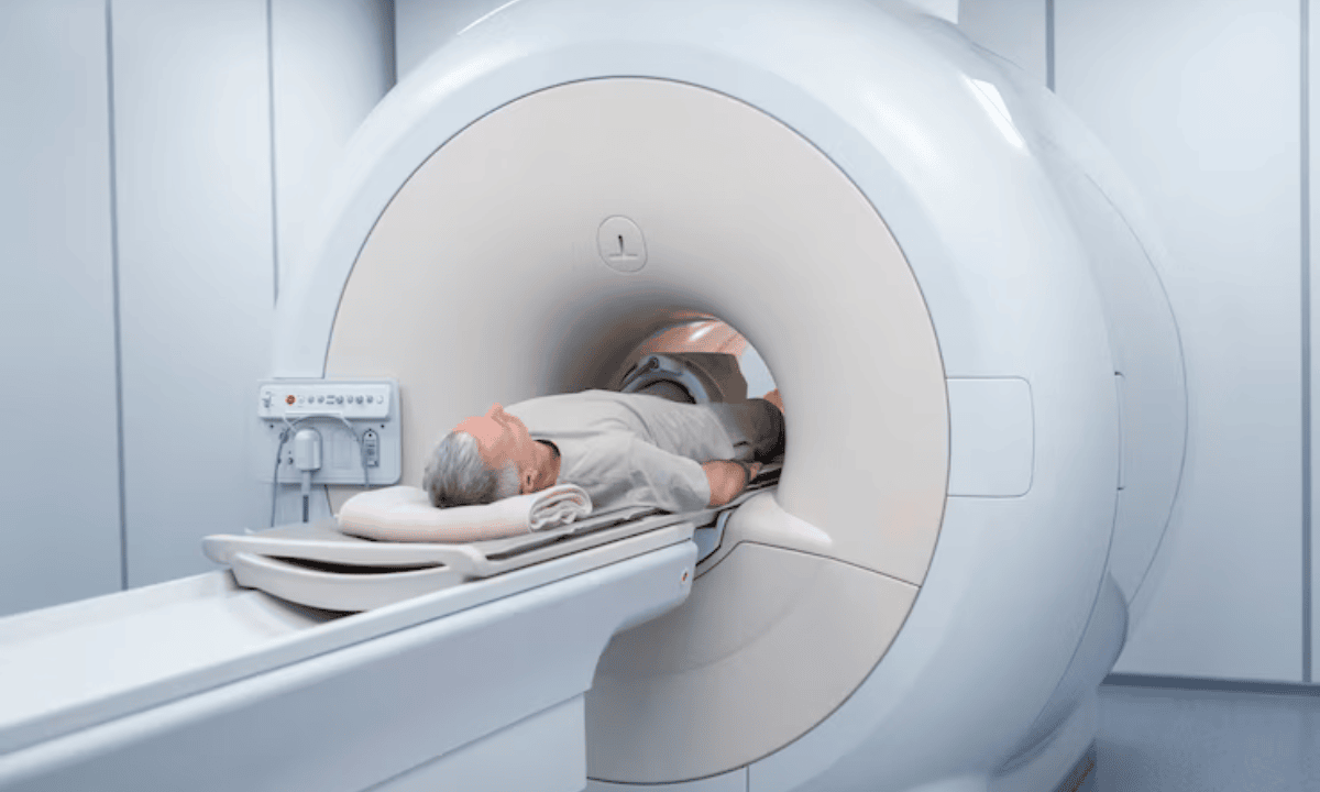
What is an MRI?
An MRI (Magnetic Resonance Imaging) is a medical imaging test that creates detailed images of your body’s tissues, organs, and bones. Your doctor may recommend an MRI to help diagnose conditions such as brain and spinal cord disorders, joint and soft tissue injuries (like ligament or tendon tears), brain injuries, tumours or abnormal growths, and diseases of your internal organs.
How to prepare for your MRI?
Wear metal-free clothing:
Choose clothing without zippers, buttons, or metal fasteners and remove all jewellery, piercings, and accessories before your appointment.
Inform your doctor about implants or metal:
Let your healthcare provider know if you have any medical implants, pacemakers, or metal objects in your body, as these may affect your safety during the scan.
Follow your usual routine:
Unless your doctor advises otherwise, eat normally and continue taking your regular medications.
Claustrophobia:
If you are uncomfortable about being in tight spaces, don’t hesitate to discuss it with your doctor before your appointment. They may prescribe you a mild sedative before the procedure to help you relax during the scan.
What is an MRA?
An MRA (Magnetic Resonance Angiography) is a specialised type of MRI scan focused on examining your blood vessels and circulation. This scan helps your doctor detect problems like narrowed or blocked arteries (atherosclerosis), aneurysms (bulges caused by weakened areas in blood vessel walls that can burst), blood clots, vascular malformations, and signs of stroke.
How to prepare for your MRA?
Follow MRI preparation guidelines:
The steps for MRA are similar to those for MRI, wear metal-free clothing, inform your doctor about implants or metal, and maintain your usual routine unless instructed otherwise.
Contrast dye considerations:
If your MRA requires contrast dye, tell your doctor if you have kidney disease or a history of allergies, as this may affect your eligibility for the procedure.
Special instructions:
For certain MRA exams (such as those involving the heart), you may be asked to avoid caffeine before the scan.
How are MRAs and MRIs similar?
You'll find that both MRA and MRI scans share several important features that make them safe and effective diagnostic tools.
They use the same technology:
Both tests rely on powerful magnets and radio waves to create detailed pictures of your body. This means you won't be exposed to harmful radiation like you would with X-rays or CT scans.
You'll have a comfortable experience:
Neither test involves needles, surgery, or any procedures that break your skin. You simply lie on a table that slides into the scanning machine.
Most patients find the process painless, though you might hear some loud knocking sounds during the scan.
They may use contrast dye:
Your doctor might give you a special dye called gadolinium through an IV before your scan. This dye helps create clearer, more detailed images.
The dye is safe for most people and helps your medical team see exactly what they need to diagnose your condition.

How are MRAs and MRIs different?
While these scans use similar technology, they focus on different parts of your body and serve distinct purposes.
MRI examines your body's structures:
When you get an MRI scan, the scan creates detailed pictures of your soft tissues, bones, and organs. Your doctor might order an MRI to look at your brain, spine, joints, or organs in your abdomen.
This test helps identify problems, like torn ligaments, brain tumours, or spine issues.
MRA focuses on your blood vessels:
An MRA specifically examines your blood vessels and how blood flows through them. This test is particularly useful for checking the blood vessels in your brain, heart, neck, and kidneys.
Your doctor might recommend an MRA if they're concerned about blocked arteries, aneurysms, or other blood vessel problems.
They handle contrast dye differently:
MRA scans often require contrast dye to clearly show blood flow patterns and highlight your blood vessels. MRI scans sometimes use contrast agents, but not always, it depends on what your doctor needs to see.
MRA vs. MRI: uses for each procedure
While both MRI and MRA use magnetic fields and radio waves to create detailed images of your body structure, they target different aspects of the body.
When is MRI used?
MRI is most useful for examining:
Brain and spine conditions:
Tumours, multiple sclerosis, or herniated discs
Joint and musculoskeletal injuries:
Torn ligaments, cartilage damage, or soft tissue injuries
Diseases of internal organs:
Liver, kidney, or heart abnormalities
When is MRA used?
MRA is useful for viewing blood vessels and is best when used for:
Stroke and aneurysm detection:
Evaluating brain arteries and carotid arteries and identifying blockages
Heart and vascular issues:
Diagnosing coronary artery disease, aortic aneurysms, or vessel narrowing
Circulatory problems in kidneys or limbs:
Detecting artery blockages in the renal arteries or leg vessels (peripheral artery disease)
For more information about MRI and MRA scans and to determine which one is most suitable for your condition, don’t hesitate to speak with a doctor. You may contact Thomson Medical to arrange a consultation for personalised guidance tailored to your individual healthcare needs.
MRI vs. MRA results
MRI and MRA are imaging tests that use magnetic fields and radio waves, but they focus on different parts of the body. Knowing what each test shows helps guide diagnosis and treatment.
What does an MRA show?
When you get an MRA, the images reveal important details about your blood vessels and circulation.
You'll see how blood flows through your brain, heart, and limbs. This helps your doctor understand if blood is reaching all the areas of your body that need it.
The scan can detect narrowed arteries or complete blockages. These problems can reduce blood flow and cause serious health issues if left untreated.
Your MRA can also identify brain aneurysms, which are areas where blood vessel walls have become weak and bulged out. Finding these early helps prevent dangerous complications.
Sometimes the scan reveals abnormal blood vessel formations that you were born with or that developed over time. Your doctor can determine if these need treatment.
What does an MRI show?
Your MRI provides detailed pictures of your body's solid structures and can reveal various types of problems.
The scan is good at showing soft tissue injuries in your ligaments, muscles, and tendons. If you've had a sports injury or accident, MRI can pinpoint exactly what's damaged.
For brain and spinal cord conditions, MRI creates clear images that help your doctor diagnose neurological problems and plan appropriate treatment. It can also detect tumours, cysts, or abnormalities in your organs. Early detection often leads to better treatment outcomes and peace of mind.
MRI also shows joint and bone diseases, helping your doctor understand arthritis, fractures, or other skeletal problems you might be experiencing. Understanding what each test shows helps you better discuss your results with your healthcare team and make informed decisions about your care.
Are there any risks with MRI or MRA scans?
Both MRI and MRA scans are generally very safe. However, there are a few things to keep in mind:
Magnetic sensitivity:
The strong magnetic field may affect your metal implants, pacemakers, or other medical devices. Always inform your doctor or radiologist beforehand.
Contrast dye side effects:
If your scan involves contrast dye, mild reactions like nausea, headache, or a rash can occasionally occur. Serious allergic reactions are rare.
Feeling confined:
Some people may feel anxious or claustrophobic inside the MRI machine. If this is a concern, let your doctor know, they may prescribe you a sedative to help calm you down during the scan.
FAQ
Which is better, MRI or MRA?
It depends on what your doctor is looking for:
MRI is ideal for detailed images of soft tissues, including the brain, spine, joints, and internal organs. It’s commonly used to detect tumours, injuries, or inflammation.
MRA (Magnetic Resonance Angiography) is a special type of MRI that focuses on blood vessels. It’s best for identifying blockages, aneurysms, or other issues affecting blood flow.
If your doctor suspects a problem with blood flow or arteries, an MRA is better. If they need a detailed look at soft tissues or brain structures, an MRI is the better choice.
What can an MRA detect that an MRI cannot?
An MRA specifically detects blood vessel abnormalities, such as:
Narrowed or blocked arteries:
Helps diagnose conditions like atherosclerosis.
Aneurysms:
Detects weak spots in blood vessel walls that could rupture.
Abnormal blood vessel formations:
Helps diagnose vascular malformations.
Blood clots:
Identifies issues like deep vein thrombosis (DVT) or embolisms.
Will an MRA show a brain tumour?
MRA typically does not detect brain tumours because it focuses on blood vessels rather than soft tissues. A regular MRI scan, especially one with contrast dye, works much better for finding brain tumours, cysts, or inflammation.
Your doctor will choose MRI when they suspect these types of problems. However, if a tumour affects your blood vessels, MRA might show abnormal blood flow patterns around the tumour area. In these cases, your doctor might order both tests to get complete information about your condition.
Will an MRA show a stroke?
Yes, an MRA is one of the best tests for detecting strokes, especially ischaemic strokes (caused by blocked arteries). It can also show:
Narrowed or blocked arteries in the brain (a major stroke risk)
Aneurysms or blood clots that could lead to a stroke
Blood flow issues that may indicate a stroke has already occurred
Why would a neurologist order an MRA or an MRI?
A neurologist may order:
MRI if they suspect:
Brain tumours, multiple sclerosis (MS), epilepsy, or nerve damage
Spinal cord injuries or herniated discs causing nerve pain
Brain inflammation or infections like meningitis
MRA if they suspect:
Stroke or aneurysm risk (narrowed or blocked blood vessels)
Carotid artery disease (blood flow problems in the neck)
Vascular malformations (abnormal blood vessel connections)
How to choose between MRI and MRA?
Choosing between MRI and MRA depends on your doctor’s diagnostic needs. MRI is best for imaging soft tissues, organs, and bones; it helps diagnose tumours, joint injuries, or brain abnormalities.
MRA, a specialised MRI, focuses on blood vessels to detect blockages, aneurysms, or circulatory issues. Your doctor will recommend the scan that best targets your symptoms
The information provided is intended for general guidance only and should not be considered medical advice. For personalised recommendations based on your medical conditions, arrange a consultation with Thomson Medical.
For more information, contact us:
Thomson Medical Concierge
- 8.30am - 5.30pm
- WhatsApp: 9147 2051
Need help finding the right specialist or booking for a group?
Our Medical Concierge is here to help you. Simply fill in our form, and we'll check and connect you with the right specialist promptly.
Notice:
The range of services may vary between Thomson clinic locations. Please contact your preferred branch directly to enquire about the current availability.
Get In Touch