
What is a PET scan?
PET (Positron Emission Tomography) is a special type of medical scan that shows how your organs and tissues are working inside your body. Unlike other medical imaging scans that just take pictures, a PET scan can show how your tissues and organs are functioning in real time.
This type of scan is particularly effective at spotting problems early because it can detect when cells start behaving differently, even before any physical changes happen that you could see on other types of scans.
What happens during a PET scan?
A PET scan shows how well your organs and tissues are working inside your body. Here's what you can expect during your procedure:
Before your scan
Your medical team will give you a small injection of a radiotracer, which is a safe radioactive material to highlight areas of increased metabolic activity. This injection is usually administered into a vein in your arm, similar to getting your blood drawn.
How does the radiotracer work?
Once injected, the radiotracer travels through your bloodstream to different parts of your body. It naturally collects in areas that are very active, such as:
Cancer cells (which are often more active than normal cells)
Inflamed or infected tissues
Parts of your brain or heart that are more active than usual
During your scan
The PET scanner can detect the tiny amounts of radiation from the radiotracer and create detailed pictures showing where it has collected. This helps your healthcare providers see how your organs are functioning.
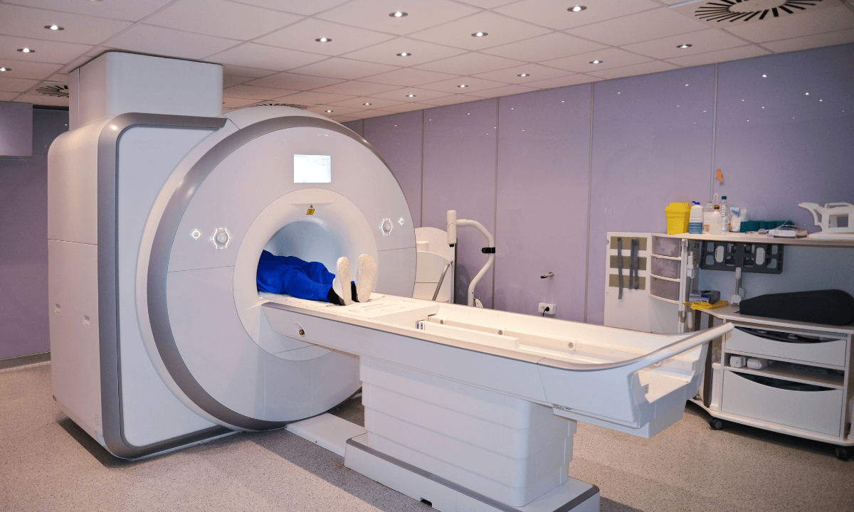
The scan usually takes between 30 minutes and 1 hour. During the scan, you’ll lie still on a flat table that slides into the PET scanner, a large, tube-shaped machine that looks similar to an MRI machine. The procedure is painless, and you won’t feel the radiotracer working in your body either.
What is an MRI?
MRI (Magnetic Resonance Imaging) scans use powerful magnets and radio waves to create very detailed pictures of the inside of your body. Think of it as taking a clear photographs of your organs, muscles, and other soft tissues without having to make any cuts or use radiation.
MRI is excellent at showing the structure and shape of things inside your body with amazing detail. It can show doctors exactly what your tissues look like and if there are any problems with their shape or size.
What happens during an MRI?
An MRI creates very detailed pictures of your body's structure using magnets and radio waves. Most MRI scans do not require injections or radiation.
How does MRI work?
The MRI machine uses powerful magnets and radio waves to take cross-sectional pictures of your body. Think of it like taking many thin slices of bread from a loaf, each "slice" shows a different layer of your body in detail.
During your scan
You'll lie on a table that slides into a large, tube-shaped machine. The machine makes loud knocking and tapping sounds, but this is completely normal. Depending on the area under scan, the procedure can take anywhere from 15 minutes to 1 hour. During the scan:
You'll need to lie very still during the imaging scan
You will be given earplugs or headphones because the machine is noisy
Sometimes you might need a contrast injection to make certain areas show up better
PET Scan vs. MRI: What's the Difference?
| Aspect | PET scan | MRI |
|---|---|---|
| Imaging type | Functional imaging, used to show how tissues and organs function | Structural imaging, used to show anatomical structure and appearance |
| Mechanism | Uses radioactive material (radiotracer) injected into the body; detects gamma rays produced when positrons interact with electrons | Uses powerful magnetic fields and radiofrequency waves |
| Radiation | Uses radiation (radiotracer) | No radiation used |
| Primary strength | Shows metabolic processes and cellular activity | Provides high-resolution images of anatomical structures |
| Best for visualising | Metabolic activity, cellular function, early disease detection | Soft tissues (brain, spinal cord, muscles, ligaments, internal organs) |
| Common medical uses | • Oncology (cancer detection) • Cardiology (heart function) • Neurology (brain activity) | • Neurological disorders • Musculoskeletal conditions • Tumour visualisation |
| Key advantage | Can identify diseases at earliest stages, even before structural changes occur | Ideal for detailed visualisation of soft tissues and anatomical structures |
| Information provided | Functional and metabolic information | Structural and anatomical information |
| Limitation | Less detailed structural information | Does not show metabolic activity or tissue function |
What is more accurate: an MRI or a PET scan?
Both MRI and PET scans are an accurate medical imaging, but they look for different things in your body. Think of it like this, one is better at taking detailed pictures, while the other is better at showing how things work.
How do PET scans work?
A PET scan looks at how active the cells in your body are. It can spot problems early because unhealthy cells often become more active before they change shape.
PET scans can show:
Cancer cells before they form visible tumors
Areas where cancer might be spreading
Brain changes in memory problems like Alzheimer's disease
Heart muscle damage after a heart attack
The main strength of PET scans is catching diseases when cells start acting differently, even before you can see physical changes.
How do MRI scans work?
An MRI takes a detailed picture of the inside of your body. It's like having a high-quality camera that can see through your skin to show muscles, organs, and other soft tissues.
MRI scans can show:
Exact size and location of brain tumors
Spine and nerve injuries
Torn muscles, tendons, or ligaments
Detailed views of organs and soft tissues
MRI provides doctors with detailed images of your body's structure, but it does not reveal the active functioning of those parts.
Which one should you have?
Your doctor will choose the right scan based on what they need to learn about your health:
PET scan:
When they need to see how your body is functioning or if a disease is active.
MRI scan:
When they need detailed pictures of specific body parts.
When do you need both?
Your doctor might recommend both PET-MRI scans to get the full picture. This combination of scans gives them two types of important information:
From PET:
How well is your body working?
From MRI:
What does your body look like inside?
Having both pieces of information helps your medical team make the most accurate diagnosis and create the best treatment plan for you.
FAQ
What does a PET scan provide that an MRI does not?
A PET scan shows functional and metabolic activity within tissues. It can reveal how tissues function at a cellular level. This level of detail is important for catching early signs of diseases like cancer before any structural changes happen. For example, PET scans can spot active cancer cells that are too small for MRI or CT scans to detect.
MRI, on the other hand, focuses on providing detailed anatomical images of organs. An MRI can show structural abnormalities, such as the presence of tumours or inflammation. However, it does not provide any information about how these tissues are functioning.
Why would a doctor recommend a PET scan?
A doctor may recommend a PET scan for various reasons, including:
Cancer diagnosis and monitoring:
This type of scan is commonly used to detect the presence of cancer, determine its location, assess whether it has spread to other parts of the body (metastasis), and monitor the effectiveness of cancer treatments like chemotherapy or radiation.
Neurological conditions:
This type of scan can help diagnose and monitor medical conditions like Alzheimer's disease, epilepsy, and brain tumours by detecting abnormal brain activity and metabolic changes.
Cardiovascular issues:
This scan is useful in evaluating heart function, identifying areas of reduced blood flow, and assessing myocardial viability in patients with coronary artery disease.
Infection and inflammation:
This scan can also detect areas of infection or inflammation in the body by highlighting regions with increased metabolic activity.
What is the biggest disadvantage of using a PET scan?
The main disadvantage is exposure to radiation from the radioactive tracer used in the imaging process. While the dose is generally low and safe, it may be a concern for pregnant women or those needing repeated medical tests.
Additionally, PET scans are more expensive and less widely available than other imaging techniques like MRIs or CT scans.
Another limitation is that PET scans do not provide as detailed images of organs or blood vessels as MRI or CT scans, so they may miss small structural changes in tissues
Does a PET scan show all cancers?
While PET scans are excellent at detecting many types of cancer, they do not show all tumours. Some tumours, including neuroendocrine tumours, don’t show up well on PET scans because they have lower metabolic activity. Furthermore, PET scans may not detect very small cancers or those in low blood flow areas, such as some brain tumours.
Moreover, inflammatory conditions or infections can sometimes cause false-positive results, meaning the scan may indicate abnormal metabolic activity in areas that are not cancerous.
In clinical practice, PET scans are often used together with other types of imaging tests like MRI, CT scans, or biopsies as part of a combination of scans for a more complete diagnostic process.
Can a PET scan and MRI be done together, and what are the benefits?
Yes, a PET-MRI scan combines both technologies into a single imaging process. This combination of scans allows doctors to get detailed images of organs and tissues (from MRI) along with information about cellular activity and metabolism (from PET).
This approach can improve the accuracy of diagnosis for certain medical conditions, such as cancer or neurological disorders, by providing both structural and functional information at the same time.
Your healthcare provider will advise if a PET-MRI scan is suitable for your situation.
Are PET and MRI scans painful or uncomfortable?
PET and MRI scans are not painful, but some people may find them uncomfortable. PET scans involve a brief injection, which can cause mild discomfort. MRI scans require lying still in a confined space for 30–60 minutes, which may be challenging for those with claustrophobia or anxiety. Both procedures are generally well-tolerated, and staff provide support for comfort.
The information provided is intended for general guidance only and should not be considered medical advice. For personalised recommendations based on your medical conditions, request an appointment with Thomson Medical.
For more information, contact us:
Thomson Medical Concierge
- 8.30am - 5.30pm
- WhatsApp: 9147 2051
Need help finding the right specialist or booking for a group?
Our Medical Concierge is here to help you. Simply fill in our form, and we'll check and connect you with the right specialist promptly.
Notice:
The range of services may vary between Thomson clinic locations. Please contact your preferred branch directly to enquire about the current availability.
Get In Touch