
What is a shoulder MRI?
A shoulder MRI (magnetic resonance imaging) is a safe and painless test that uses a magnetic field and radio waves to produce detailed pictures of the inside of your shoulder. The test examines the soft tissues around the shoulder joint, such as muscles, tendons, ligaments, cartilage, and nerves.
While X-rays or CT scans concentrate on the bones, an MRI is especially suited for imaging soft tissue injuries or conditions. No radiation is used in an MRI, which is why it is a safer alternative for certain conditions, such as in younger patients and those receiving multiple scans.
When is a shoulder MRI scan used?
A shoulder MRI is typically used when a physician needs more detailed information about your shoulder joint and surrounding tissues. It’s often ordered when:
Rotator cuff injuries:
MRIs can diagnose partial or full-thickness rotator cuff tears, a common condition among athletes, labourers, and older individuals.
Labral tears:
The labrum is a cartilage ring that helps stabilise your shoulders. MRI scans are particularly effective in detecting labrum tears, which can occur due to dislocations or repeated stress.
Shoulder impingement syndrome:
This condition occurs when the tendons of the rotator cuff become irritated or compressed. MRI is valuable in identifying the inflammation and soft tissue damage associated with impingement.
Arthritis:
MRI can show early signs of joint degeneration (arthritis), such as cartilage thinning or bone spurs, that may not be visible on X-rays.
Bursitis and tendinitis:
MRI is excellent for identifying inflammation in the shoulder’s bursa or tendons.
Fractures or bone abnormalities:
X-rays are the first step to diagnose broken bones (fractures). But MRI can spot stress fractures or bone contusions that regular X-rays may not reveal.
Infections or tumours:
MRI can find tumours, cysts, or infections in the soft tissues of the shoulder. This finding may require different treatment options.
Frozen shoulder (adhesive capsulitis):
MRI can help to assess frozen shoulder (adhesive capsulitis) and the extent of joint stiffness and check if other conditions are causing symptoms.
For more information about shoulder MRI scans and to determine if it’s appropriate for your condition, consider speaking with a healthcare professional. You may contact Thomson Medical to arrange a consultation for personalised guidance tailored to your individual care needs.
How do I prepare for a shoulder MRI?
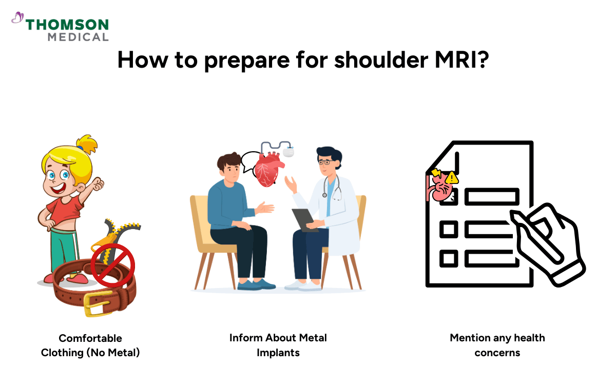
Preparing for your shoulder MRI is usually simple, but there are a few important steps you'll need to follow:
Discuss your medical history with your doctor:
Inform your doctor if you have any metal implants or devices, such as pacemakers, artificial joints, stents, artificial heart valves, or surgical clips, as these can be affected by the magnetic field.
Certain devices, like some aneurysm clips, may prevent you from undergoing an MRI.
Avoid wearing metal items:
Before your imaging tests, remove all jewellery, watches, piercings, or other metallic objects, as these can interfere with the magnetic field.
Wear comfortable clothing:
It’s best to wear loose-fitting, comfortable clothes.
If your clothes contain metal objects or if you're receiving an injection of contrast material, your doctor might ask you to change into a hospital gown.
Inform about any claustrophobia or anxiety:
MRI machines can be enclosed, which can cause anxiety or discomfort for some patients.
If you are claustrophobic, talk to your doctor, as they may offer sedation or refer you to a facility that offers an open MRI, which has a larger opening.
Contrast material:
If your doctor has ordered an MRI with contrast (usually gadolinium-based dye), you will receive an injection into your vein before or during the procedure.
Make sure to inform the technician if you have an allergy to gadolinium (contrast agents), kidney problems, or a history of nephrogenic systemic fibrosis (NSF).
How does the test work?
Your MRI works by using a powerful magnet to create a magnetic field around your body. The machine sends out radiofrequency pulses that temporarily change the alignment of hydrogen atoms in your body.
When these pulses stop, the atoms return to their normal position and release energy. The MRI machine's sensors pick up this energy, and a computer processes it to create highly detailed images of the structures inside your shoulder.
Your scan can show not only your bones but also soft tissue details like tendons, ligaments, muscles, nerves, and cartilage. This detailed view is especially helpful for diagnosing shoulder problems that other tests might miss.
What to expect during your scan?
Your procedure will typically take 30 to 60 minutes. You'll lie still on a table, and your affected shoulder will be positioned inside the MRI machine. If your doctor ordered contrast dye, it may be injected into your shoulder joint or through a vein in your arm to make the images clearer.
The procedure itself is non-invasive and usually painless. However, the machine can be quite loud during the scan, so you'll likely be given earplugs or headphones to reduce the noise and help you feel more comfortable.
What are the potential risks and side effects of this test?
MRI scans are considered safe, with minimal risks involved. However, there are a few potential risks and side effects:
Claustrophobia:
The MRI machine is a narrow, enclosed tube, which may cause anxiety or discomfort in some individuals. In such cases, a sedative may be given before the procedure, or an open MRI may be offered.
Contrast reaction:
While rare, some patients may experience an allergic reaction to the contrast dye (gadolinium), which could result in itching, rash, or, in severe cases, difficulty breathing.
Kidney patients are at higher risk for complications related to contrast dye.
Metal implants or devices:
People with certain metal implants (such as pacemakers, cochlear implants, or metal surgical clips) may not be able to undergo an MRI due to the powerful magnetic field.
It’s essential to inform your doctor and MRI technician about any implants.
Discomfort from lying still:
You may need to lie still for 30 to 60 minutes, which can be uncomfortable, especially if you already have shoulder pain.
Pregnancy considerations:
While MRIs are generally considered safe during pregnancy, doctors typically avoid them during the first trimester unless absolutely necessary.
Always inform the technician if you are pregnant or suspect you might be.
Loud noises:
The MRI machine makes loud tapping or thumping sounds during the scan. Usually, the technician will give you earplugs or headphones to reduce the noise.
FAQ
What does an MRI of the shoulder show?
A shoulder MRI provides detailed, high-resolution images of the bones, muscles, tendons, ligaments, cartilage, and nerves in your shoulder.
This imaging method helps your healthcare provider assess a wide range of conditions, including soft tissue injuries like rotator cuff or labral tears, joint disorders such as arthritis, and inflammation from bursitis or tendinitis.
It can also reveal more serious medical conditions, such as infections, cysts, or tumours, giving your doctor the information needed for an accurate diagnosis and effective non-invasive treatments.
How long does a shoulder MRI usually take?
The entire procedure typically lasts between 30 and 60 minutes. Your health care provider will let you know if contrast material is needed, which may extend the scan time. If you're undergoing a more complex medical examination with additional views, it could take slightly longer.
Is a shoulder MRI open or closed?
A closed MRI machine, a tunnel-like scanner that provides high-quality images, performs most shoulder MRIs. If you are anxious about enclosed spaces, some clinics may offer open MRI machines with a wider design.
While open scanners can be more comfortable, especially for those with mobility concerns or claustrophobia, they may not always produce MRI images with the same level of detail as closed systems.
Can MRI show nerve damage in the shoulder?
An MRI cannot directly test nerve function, but it can provide a detailed diagnosis by revealing structural changes that suggest nerve involvement. For example, MRI images can show if a rotator cuff tear, arthritis, or a herniated disc is pressing on nearby nerves.
If your doctor suspects nerve damage, they may also recommend additional tests, such as nerve conduction studies or electromyography (EMG), to support the medical diagnosis.
Which is better, an MRI or a CT scan for the shoulder?
The type of condition under examination determines the choice between an MRI and a CT scan.
MRI is generally better for assessing soft tissues like muscles, tendons, ligaments, and cartilage, making it the preferred method for diagnosing injuries like rotator cuff disorders, labral tears, and arthritis. CT scans, on the other hand, are more effective at evaluating bone fractures and structural issues in hard tissues.
For most shoulder problems, an MRI is often the preferred medical imaging technique, but CT scans may be ordered if there is concern about bone fractures or complex joint issues.
What is the difference between a normal and an abnormal shoulder MRI?
Your MRI imaging exam of the shoulder has shown that there are no problems visible. If the results are abnormal, this means that the scans have shown problems with your shoulder. The cause could be anything from a tear to arthritis to a cyst. Your doctor will explain your results and what to do next.
The information provided is intended for general guidance only and should not be considered medical advice. For personalised recommendations based on your medical conditions, arrange a consultation with Thomson Medical
For more information, contact us:
Thomson Medical Concierge
- 8.30am - 5.30pm
- WhatsApp: 9147 2051
Need help finding the right specialist or booking for a group?
Our Medical Concierge is here to help you. Simply fill in our form, and we'll check and connect you with the right specialist promptly.
Notice:
The range of services may vary between Thomson clinic locations. Please contact your preferred branch directly to enquire about the current availability.
Get In Touch