
Receiving a diagnosis of stage 0 breast cancer can be worrying, and it's completely natural to feel overwhelmed and have questions about what it means for your health. However, an early diagnosis gives you time to make an informed decision about your treatment.
Knowing what stage 0 means, what treatment options are available and what to expect along the way can help you feel more in control as you navigate your care.
What is stage 0 breast cancer?
Stage 0 breast cancer is the earliest stage of breast cancer. It means abnormal cells are found in the lining of the breast milk ducts. The cells have not spread into nearby breast tissue or spread elsewhere in the body. Your doctor may call this carcinoma in situ, which means the cells are still in the place where they first developed.
Because the cells have not invaded surrounding tissue, stage 0 is described as non-invasive. Without treatment, these abnormal cells can develop into invasive breast cancer over time. That is why some form of treatment is usually recommended, even if you feel completely well.
What are the types of stage 0 breast cancer?
While stage 0 is non-invasive, it can look different in each person. Identifying the type helps your doctor to recommend suitable treatment options for you.
Ductal carcinoma in situ (DCIS)
DCIS is the most common form of stage 0 breast cancer. In this condition, abnormal cancer cells are found inside the milk ducts but have not spread to nearby breast tissue, lymph nodes, or other parts of the body.
Paget’s disease of the nipple
This is a rare form of stage 0 breast cancer where cancer cells are present in the skin of the nipple or the darker area around it.
You might also come across lobular carcinoma in situ (LCIS). Despite its name, LCIS is not breast cancer. It's a breast change that may raise your risk of developing breast cancer in the future. LCIS doesn't usually require treatment, but closer monitoring, such as regular mammograms, may be recommended for you.
Does stage 0 breast cancer have symptoms?
Stage 0 breast cancer often doesn't cause any symptoms. It's usually picked up during your routine mammography screening, before you notice any changes.
In some cases, you might notice:
A noticeable lump in the breast
Unusual nipple discharge, including blood
Whether your diagnosis came as a complete surprise or followed a symptom you noticed, understanding how stage 0 is diagnosed can help you feel more prepared for what comes next.
If you have noticed any breast changes or have questions about your screening, speaking with a healthcare provider is a good first step. Request an appointment with Thomson Breast Centre, where our specialists can help to determine what further assessment, if any, may be right for you.
Our breast cancer specialists in Singapore
Loading...
How is stage 0 breast cancer diagnosed?
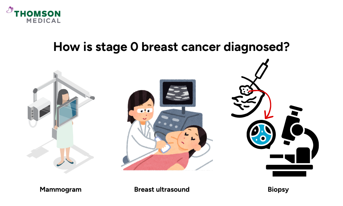
Hearing that you need further tests can feel unsettling, but diagnosis for stage 0 breast cancer is straightforward and designed to give you a clearer picture of your health.
Mammogram
A mammogram is a low-dose breast X-ray. It's often how stage 0 breast cancer is first spotted. During the scan, your breast is gently compressed between two plates for a few seconds to produce clear images of the tissue.
One of the common findings on a mammogram is breast calcifications (also called calcium deposits), which appear as white flecks or white spots on the image. If something like this is seen, your doctor may ask you to return for a diagnostic mammogram or other imaging tests for a closer look.
Breast ultrasound
A breast ultrasound uses sound waves to create detailed images of your breast. If a lump is found, it helps your doctor determine whether it's a fluid-filled cyst, which is usually not cancerous, or a solid area that needs further assessment.
Your doctor may also recommend an ultrasound alongside your mammogram if you have dense breasts. Dense breasts have more tissue and less fat, which can make it harder to see abnormal areas on a mammogram alone.
Biopsy and grading
If imaging shows an abnormal finding, your doctor will perform a breast biopsy. A small sample of cells is taken from the area and examined under a microscope. The results are included in your report, which explains what the cells look like and whether they are cancerous.
When cancer cells are found, your report also notes their grade from 1 to 3. The grade is separate from the stage. It shows how the cells look compared to normal breast cells and how quickly they are likely to grow.
The grade describes:
| Grade | What it means |
|---|---|
Grade 1 (low-grade DCIS) | Cells look similar to healthy breast cells and grow slowly |
Grade 2 (moderate-grade DCIS) | Cells grow at a moderate pace |
Grade 3 (high-grade DCIS) | Cells look quite different from healthy cells and grow more quickly |
The grade is not a prediction of what will happen to you. It helps your doctor choose the most suitable treatment for you.
For example, if your DCIS is high-grade, it tends to grow more quickly, so your treatment may include radiotherapy after surgery to reduce the risk of the cancer returning. If it's low-grade, it grows more slowly and may not always need additional treatment.
Genomic and genetic testing
There are also genomic tests that analyse a sample of your DCIS to see how active certain genes are. This gives your doctor a fuller picture of how the cancer cells are likely to behave and helps personalise your treatment plan.
Depending on your health and family history, your doctor may also discuss genetic testing with you. This can provide more information about inherited risk and help shape your longer-term care plan.
If you’ve had abnormal breast imaging, it’s important to understand your results and treatment options. Request an appointment with Thomson Breast Centre to help you understand what it means and walk you through your options.
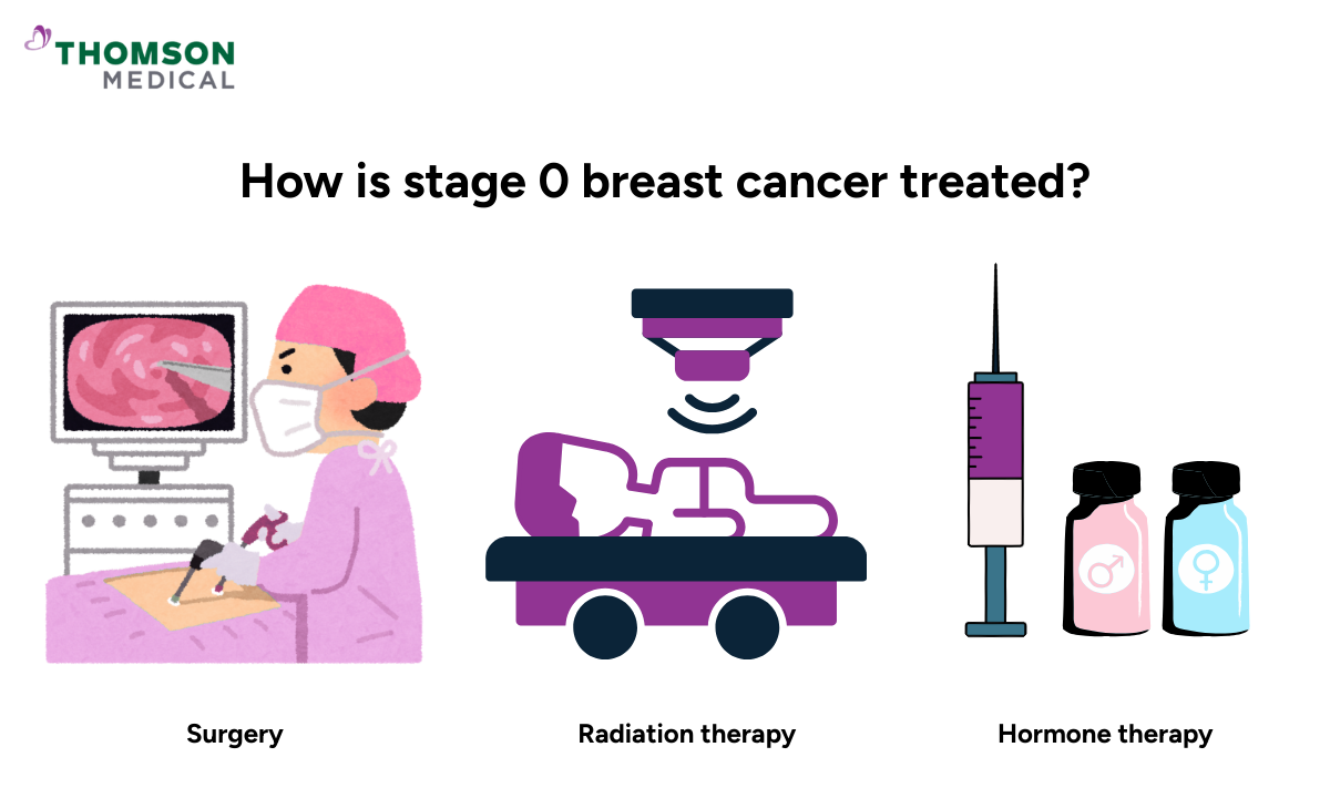
How is stage 0 breast cancer treated?
Once your diagnosis is confirmed, your doctor will work with you to decide the appropriate treatment based on your specific situation. There is no single path that works for everyone.
Your treatment will depend on:
The grade and size of your cancer cells
Your age and health history
Your personal preferences about each of the options
Take your time with this. Because stage 0 is non-invasive, your treatment decisions are usually made thoughtfully rather than urgently. That means you will have time to ask questions and weigh the side effects of each option carefully so you feel confident about the direction you choose.
Active surveillance
If you have just been diagnosed, you might assume treatment needs to start immediately. For some women, that’s not always the case.
If your cancer cells are slow-growing and low-grade (especially for older patients), your doctor may suggest active surveillance instead of immediate surgery. This doesn't mean your condition is being ignored. It means your doctor has carefully reviewed your situation and determined that close monitoring is the most appropriate path for you right now.
This means:
Regular mammograms to check for any changes
Close monitoring by your medical team
No immediate surgery unless something changes
If this option is recommended, your doctor will explain clearly why it's the most appropriate path for you. You will always have a clear plan in place, and your care team will continue to monitor for any changes.
Surgery
Surgery is the most common treatment for stage 0 breast cancer. Your surgeon will discuss which approach is the most suitable for you, but there are two main options:
Lumpectomy (the more common option):
Removes only the abnormal tissue and a small border of healthy tissue around it
Your lymph nodes are not usually removed
Research generally suggests outcomes are comparable to mastectomy for many people with stage 0 disease
Mastectomy:
This involves removal of the entire breast.
It's generally recommended if your cancer cells are present in multiple areas or the affected area is too large for a lumpectomy
If a mastectomy is needed, breast reconstruction can often be performed either at the same time or later. Your surgeon will discuss these options with you before surgery.
Radiation therapy
After a lumpectomy, your doctor may recommend radiation to lower the chance of the cancer coming back. It usually starts 3 to 4 weeks after surgery, giving your body time to heal first.
Radiation is not always needed, especially if your cancer cells are small or slow-growing. Your doctor will assess whether the benefits outweigh the risks in your case.
Hormone therapy (endocrine therapy)
If testing shows that your stage 0 breast cancer is oestrogen-receptor positive (meaning the cancer cells grow in response to the oestrogen hormone), hormone therapy may be advised after surgery.
This usually involves you taking a daily tablet for up to five years. It works by reducing the effect of oestrogen on any remaining cancer cells, lowering your risk of recurrence. The type of medication used during the therapy will depend on your menopausal status and overall health.
FAQ
Can stage 0 breast cancer come back after treatment?
Yes, there is a small risk of recurrence after treatment. High-grade DCIS (Grade 3) carries a higher chance of returning than low-grade DCIS because the cells are more aggressive and look less like normal breast cells. Lower-grade DCIS, where cells still closely resemble normal breast cells, has a lower recurrence risk.
Regular follow-up care and mammograms are important for catching any changes early.
Does family history affect my risk of stage 0 breast cancer?
Having a family history of breast cancer can increase your risk. However, many people diagnosed with stage 0 breast cancer have no family history at all.
What is comedo necrosis?
Comedo necrosis refers to areas of dead cancer cells found inside the breast milk duct. This happens when cancer cells grow so quickly that cells at the centre no longer receive enough blood supply. It's associated with high-grade DCIS and may influence your treatment plan.
What is the risk after a lumpectomy?
The risk after lumpectomy refers to the chance of stage 0 breast cancer or invasive cancer returning in the same breast. This risk is generally low but can be further reduced with radiation therapy or hormone therapy, depending on your specific situation.
Can stage 0 breast cancer affect my ability to have children?
Stage 0 breast cancer treatment doesn't directly affect fertility. However, hormone therapy such as tamoxifen is not recommended during pregnancy. If you are planning to have children, speak with your doctor before starting treatment so your treatment plan can take this into consideration.
Why is radiation recommended after a lumpectomy?
Radiation after lumpectomy helps reduce the risk of the cancer returning in the same breast. While surgery removes the visible abnormal tissue, radiation after lumpectomy would target any remaining cells that may not have been detected. Together, these treatments lower your chances of a cancer cell developing into invasive breast cancer.
The information provided is intended for general guidance only and should not be considered medical advice. For personalised recommendations and tailored advice based on your unique situations, request an appointment with Thomson Breast Centre today.
Reference:
Giannakeas, V., Irvine, M. A., Pungchanchai, N., Tao, L., & Narod, S. A. (2018). Association of radiotherapy with survival in women treated for ductal carcinoma in situ with lumpectomy or mastectomy. JAMA Network Open, 1(4), Article e182320. https://pmc.ncbi.nlm.nih.gov/articles/PMC6324271/
Wang, Y., & Xu, W. (2024). Progression from ductal carcinoma in situ to invasive breast cancer: Molecular features and clinical significance. Signal Transduction and Targeted Therapy, 9(1), Article 79. https://doi.org/10.1038/s41392-024-01779-3