
What are heart scans?
A heart scan, also called a cardiac CT (cardiac computed tomography) scan, is a special imaging test that uses X-ray technology to create detailed, high-quality images of your heart. It shows all four chambers of your heart, the heart valves, the muscle tissue, and the protective sac around the heart (called the pericardium).
Why would you need a heart scan?
Your healthcare team may recommend a heart scan for several reasons, depending on your symptoms and medical history. You may need a heart scan to:
Find the cause of symptoms like chest pain, fainting episodes (syncopal attacks), or irregular heartbeats (palpitations).
Check your heart arteries for any calcium build-up, plaque, narrowing, or blockages that could lead to heart disease.
Assess how well your heart valves are working and detect any signs of heart valve disease (valvular dysfunction).
Look at your abdominal aorta to check for issues such as aneurysms or tears (dissections).
Detect any signs of congenital heart conditions (problems you may have been born with).
Identify tumours or abnormal masses in or around your heart.
Help plan for heart surgery, valve surgery, or procedures like ablation for abnormal heart rhythms.
Monitor the progression and possible complications related to heart or heart valve surgery and ablation procedures.
This scan helps your doctor make an accurate diagnosis and choose the best treatment for your heart health.
7 common heart scans and tests
Chest x-ray
A chest X-ray is often a first step in checking your heart and lungs. It gives your doctor a brief look at the size and shape of your heart, as well as your lungs and bones. Your doctor may recommend this examination to see your heart and aorta before moving on to more detailed heart scans.
Blood test
Blood tests help your doctor learn a lot about your heart health. In an emergency, they can measure substances in the blood to show how much damage the heart muscle has suffered.
Your heart releases chemicals into your blood when it damages your muscles during a heart attack. Such substances can be measured by a blood test.
The tests are also used to check for high cholesterol and diabetes and to monitor your kidney and liver function before choosing the safest medication for your heart condition.
12-lead electrocardiogram (ECG)
12 lead ECG is often the first test done to review the electrical activity of the heart. The cardiac disorders, such as arrhythmias, thickened heart muscles, and heart attacks, will show abnormal changes on an ECG.
Your healthcare provider will attach 10 electrodes from the ECG to your arms, legs, and chest to produce 12 different views from the entire heart to represent your heart’s electrical activity.
Exercise stress test
An exercise stress test is an ECG test done while the person is exercising. The purpose of this test is to evaluate the electrical activity of the heart under stress.
Exercise helps the heart work harder and faster, which can help find abnormalities with blood flow in the heart, blood pressure fluctuations, and difficulties with heart rhythm.
The test usually requires you to walk or run on a treadmill or ride a stationary bike, during which the heart rhythm, blood pressure, and breathing are monitored.
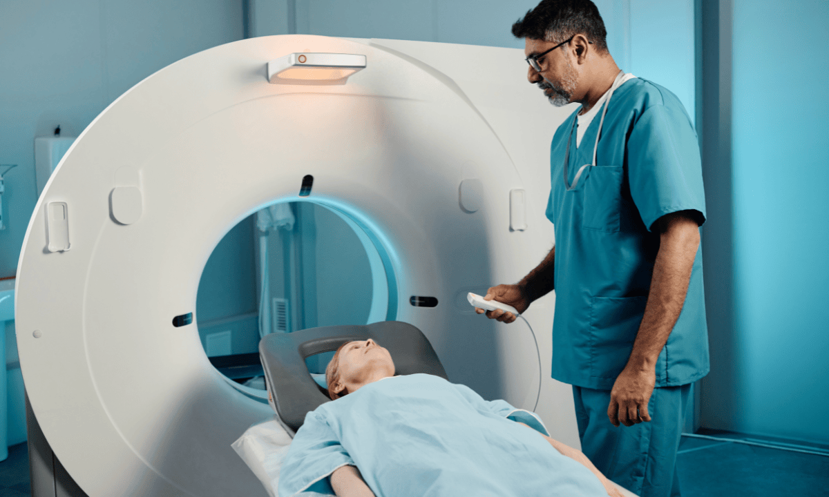
Magnetic resonance imaging (MRI)
Magnetic Resonance Imaging (MRI) is an imaging test that uses radio waves and magnetic resonance to produce a detailed image of your heart. In some cases, radiocontrast dye is required to make the parts of the heart and coronary arteries more visible.
The MRI can show the doctor the structure of the heart and how well it is functioning and if there is scarring within the heart.
Echocardiogram
An echocardiogram is an imaging test that uses ultrasound to produce clearer images of your heart. It is used to assess the structure and function of your heart.
Your doctor may suggest echocardiograms if you have symptoms such as breathlessness, leg swelling and reduced effort tolerance. It can detect the buildup of fluid within the sac surrounding the heart and identify valvular problems.
CT scan of the heart
A CT scan of the heart is an imaging test from multiple X-rays to produce a detailed image of the heart. A coronary calcium scan is a CT scan of the heart to check for hardened buildups of calcium and blood cholesterol that can cause heart attacks.
If you are at risk of heart attack, a coronary calcium scan is able to determine the risk of heart disease. Your healthcare provider may suggest additional tests or lifestyle modifications based on your risk.
For more information about heart scans and to determine if it’s appropriate for your condition, consider speaking with a healthcare professional. You may contact Thomson Medical to arrange a consultation for personalised guidance tailored to your individual healthcare needs.
What do your heart scan results mean?
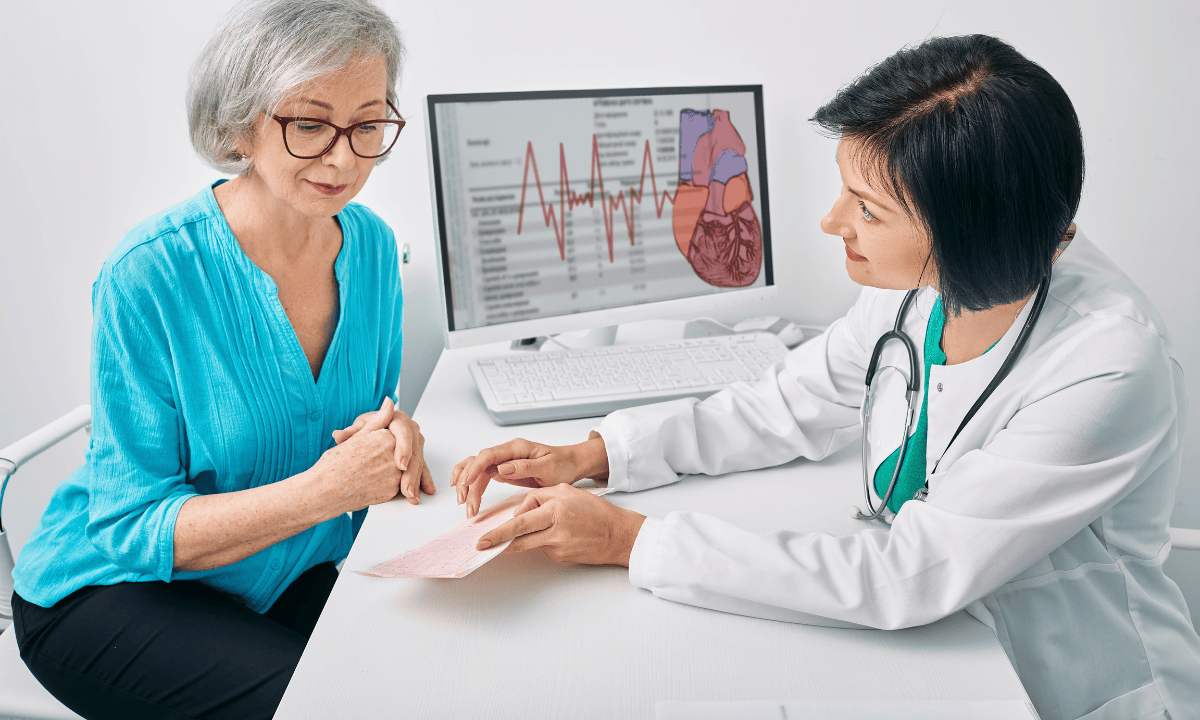
After your heart scan, your healthcare provider will go over the results with you. These will show whether everything looks normal or if there are signs of heart disease or other conditions.
Normal results
If your results are normal:
Your heart and blood vessels appear healthy with no signs of narrowing, blockages, or damage.
Your calcium score is 0, meaning no visible calcium build-up in your coronary arteries. This indicates that you do not have coronary artery disease, and your risk of a heart attack is very low.
Abnormal results
If something unusual is found, your scan may show:
Plaque or narrowing in the coronary arteries (called stenosis), which can affect blood flow to the heart.
A calcium score higher than 0, which indicates the presence of coronary artery disease:
1-100: Mild evidence
101-400: Moderate evidence
Over 400: Extensive coronary artery disease
Your report may include a percentile score, comparing your results with others of the same age, sex, and ethnicity.
Other possible abnormal findings include:
Your heart's function may be weakened, or the heart valves may not be functioning properly.
Fluid around your heart (pericardial effusion) or inflammation of your heart lining (pericarditis).
A tumour or mass is present in or around your heart.
Congenital heart abnormalities refer to conditions that are present from birth and impact the functioning of your heart.
Problems with your blood vessels, such as:
Aneurysms (enlarged areas)
Dissections (tears in the vessel wall)
Narrowing or coarctation (restricted blood flow)
Your doctor will explain what your specific results mean and help plan the next steps, whether that’s further testing, treatment, or lifestyle changes to protect your heart health.
What are the potential risks of a heart scan test?
A heart CT scan is generally safe, but like any medical test, it does have some possible risks. Here's what you should know:
Contrast dye reactions
The test uses a special dye (called a contrast agent) that contains iodine to help create clearer pictures of your heart.
Common reactions:
The following reaction may happen:
Itching
Feeling sick to your stomach
Sneezing
Skin rash
These symptoms usually go away on their own, but your medical team can give you medicine like antihistamines or steroids if needed.
Serious allergic reactions are rare, but they can occur. A more severe reaction (anaphylaxis) could cause difficulty breathing and may require emergency treatment. Be sure to tell your healthcare provider if you’ve had any allergic reactions to contrast dye in the past.
Special considerations
If you have diabetes or kidney problems:
If you have diabetes or kidney problems, your doctor may recommend that you drink extra fluids after the scan to help flush the dye from your body and protect your kidneys.
If you're breastfeeding:
If you are breastfeeding, it's possible for a small amount of the contrast dye to pass into your breast milk. You may wish to pump and store milk before the test to feed your baby for the following 24–48 hours as a precaution.
If you're pregnant:
Because X-rays can harm your developing baby, we do not recommend this test during pregnancy. If the scan is absolutely necessary, your doctor will take special steps to protect your baby.
Radiation exposure
CT scans use X-rays, which involve a small amount of radiation. While there's a minimal risk of cancer from radiation over many years, the amount used is kept as low as possible while still getting good pictures of your heart.
Heart rate medicine
Your doctor may give you medication before the scan in some cases to slow your heart rate. This helps to produce clearer images of your heart.
If you have asthma, chronic obstructive pulmonary disease (COPD), or heart failure, this medication could cause shortness of breath or worsen your symptoms.
Always inform your healthcare team if you have any of these conditions or if you’ve had trouble breathing during previous scans.
Tell your healthcare team beforehand
To avoid any risk following the scan, make sure to inform your medical team beforehand if you have:
Allergies (especially to iodine or contrast dye)
Kidney disease or liver problems
Diabetes
Asthma, heart failure, or COPD
If you're pregnant or might be pregnant
If you're breastfeeding
Remember, your healthcare team is qualified in managing these risks and will monitor you carefully throughout the procedure.
For a clearer picture of your heart health, consider our heart screening pacage. You may contact Thomson Medical to arrange a consultation for personalised guidance tailored to your individual healthcare needs.
FAQ
What does a heart scan tell you?
A heart scan provides detailed images of your entire heart, including the pumping function, heart defects, and the condition of surrounding blood vessels. It allows your doctor to assess the structure of your heart, detect calcium or plaque buildup, and check for any abnormalities such as valvular disease, aortic aneurysms, or congenital heart defects.
Who needs a heart scan?
Your doctor may suggest a heart scan for you if you are:
Having symptoms associated with a heart attack, such as chest pain, palpitations, and shortness of breath
Suspected with calcium or cholesterol build up within your blood vessels
Suspected having valvular disease
Suspected having aortic aneurysm or dissection
Suspected with heart tumour
Having congenital heart disease
How long does a heart scan take?
The entire heart scan takes around 30 to 60 minutes, including preparation time. The heart scan itself usually takes around 20 minutes.
What are the risk factors that might lead to needing a heart scan?
If you have cardiovascular disease risk factors, your doctor may recommend a heart scan for you. These factors can include a family history of heart disease, high blood pressure, high cholesterol, diabetes, smoking, obesity, or a sedentary lifestyle.
What is the best test to check for heart problems?
There is no single best test to check for heart problems. Your doctor may suggest a few tests to detect and diagnose your heart conditions based on your symptoms and risk factors. Your doctor may also offer you blood tests, electrocardiograms, echocardiograms, exercise stress tests, and cardiac MRI scans as additional tests.
Which is better, an MRI or a CT scan for the heart?
There is no single better scan for the heart. MRI heart scans focus on the internal structure and function of the heart, while CT heart scans focus on blood vessels and the detection of blockages.
CT heart scans are faster at producing images, which are often offered during emergency situations, while MRI takes longer than CT scans. A CT scan uses X-ray radiation, which is not recommended in pregnant women and children, while an MRI does not use radiation.
The information provided is intended for general guidance only and should not be considered medical advice. For personalised recommendations based on your medical conditions, request an appointment with Thomson Medical.
For more information, contact us:
Thomson Medical Concierge
- 8.30am - 5.30pm
- WhatsApp: 9147 2051
Need help finding the right specialist or booking for a group?
Our Medical Concierge is here to help you. Simply fill in our form, and we'll check and connect you with the right specialist promptly.
Notice:
The range of services may vary between Thomson clinic locations. Please contact your preferred branch directly to enquire about the current availability.
Get In Touch