
Have you sustained an injury to your knee while participating in sports? You might have a torn anterior cruciate ligament (ACL). A Magnetic Resonance Imaging (MRI) scan helps doctors to see what is happening inside your knee and how serious your ACL injury is.
Getting an accurate diagnosis helps ensure that you receive the right treatment for a full recovery. MRI scans are the most reliable way to confirm an ACL tear and assess what else might be damaged around your knee.
What is an ACL MRI?
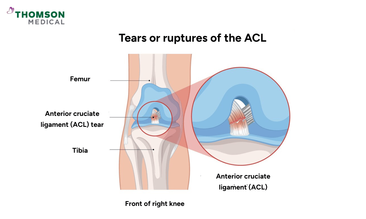
An ACL MRI is a type of knee MRI scan to visualise your anterior cruciate ligament, an important part of your knee. The ACL is one of the main ligaments that help keep your knees stable, especially during activities such as running, jumping, or pivoting.
An MRI scan uses strong magnetic fields and radio waves to produce high-resolution images of the soft tissues in your knee. It can detect the ACL, as well as surrounding cartilage, muscles, tendons, and other ligaments.
Doctors usually recommend an ACL MRI to look for tears, injuries, or other complications of the ligament and surrounding areas. It gives far more detail than an X-ray scan and is the preferred way to investigate soft tissue injuries.
When do I need a knee ACL tear MRI scan?
Your doctor might recommend an ACL MRI if they suspect an ACL injury, often after a sports-related injury or accident. Here are some conditions where this test may be recommended, which include:
Knee instability:
You may feel as though your knee is "giving way" or is unable to support your weight.
Pain:
You have persistent knee pain after an injury.
Popping sensation and swelling:
You might experience a popping sensation at the time of injury, often followed by rapid swelling in the knee.
Decreased range of motion:
You have difficulty in bending or straightening your knee.
Inability to continue sports or physical activities:
You have difficulty in performing non-linear movements that involve cutting, jumping, or pivoting.
An ACL MRI can help confirm whether the ligament is torn, partially torn, or otherwise damaged. It also helps in determining the severity of the knee injury, which is important in deciding the treatment approach.
If you're experiencing persistent knee pain, instability, or swelling—especially after a sports injury or sudden movement—it’s important to consult a healthcare provider. Request an appointment with Thomson Medical for a comprehensive evaluation.
Our specialists will assess your symptoms and may recommend a knee ACL tear MRI scan to accurately diagnose your condition and develop a personalised treatment plan tailored to your needs.
Orthopaedist in Singapore
Loading...
How do you prepare for an ACL MRI?
Preparing for an ACL MRI is simple, but there are some important steps you need to take to ensure accurate results and a smooth experience. These include:
Clothing:
Wear loose, comfortable clothing.
You may be asked to change into a hospital gown for the procedure, as metal or certain clothing materials can interfere with the MRI machine.
Metal objects:
Inform the MRI technician about any metal implants, pacemakers, joint replacements, or piercings you have, as these can interfere with the magnetic field.
Remove all jewellery, watches, credit cards, and other metal objects before the scan.
Medications:
Bring a list of all medications you’re currently taking.
In rare cases, your doctor might advise pausing some medications before the procedure.
Contrast dye:
In some cases, you may be given a contrast dye (gadolinium) to help provide clearer images of your ACL and surrounding structures.
Your healthcare team will give you specific instructions if there's anything else you need to do to prepare.
How does the test work?
The testing process
During the MRI procedure:
Before the scan, if a contrast dye is needed, it will be injected into a vein, usually in your arm. This dye helps highlight tissues and structures in your knees for better visualisation.
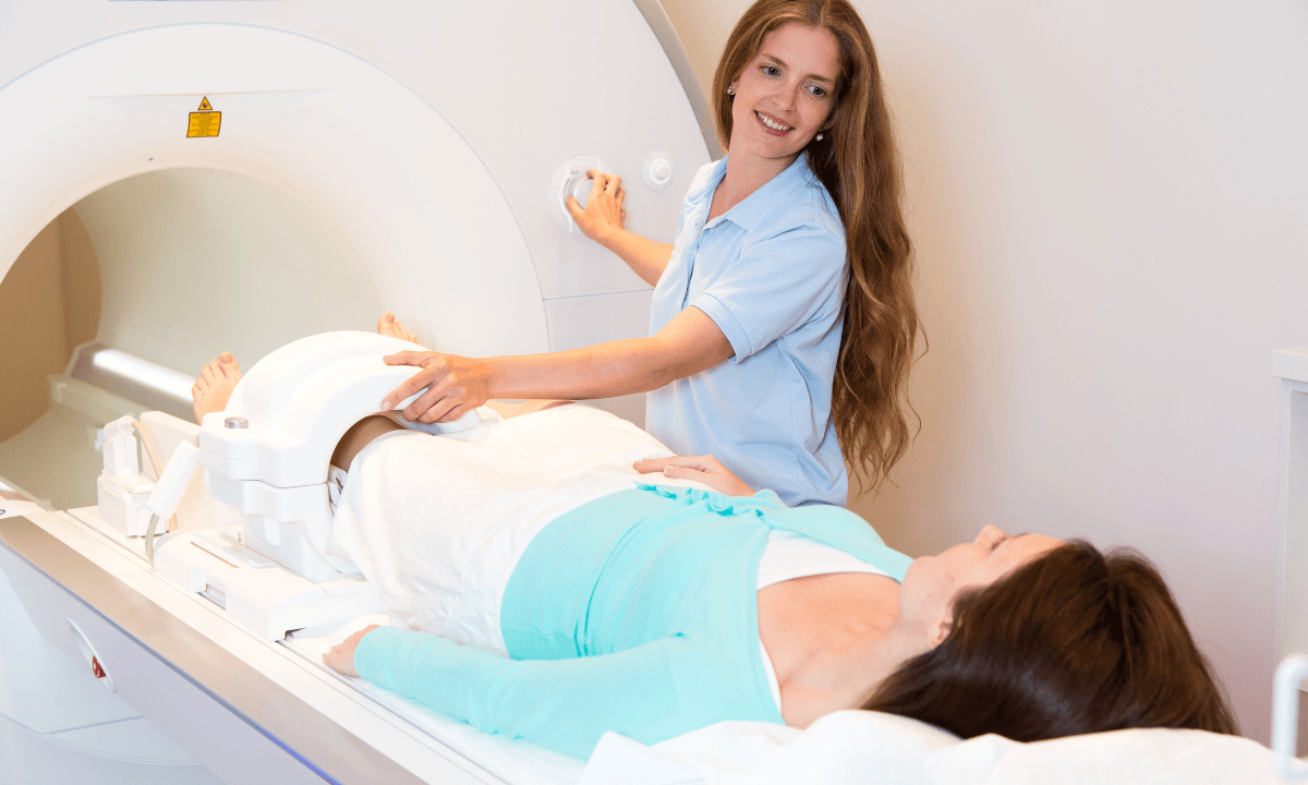
You will lie down on a narrow table that slides into the MRI machine, a large, tube-shaped device.
The machine uses strong magnetic fields and radio waves to take detailed pictures of your knee
You will need to stay as still as possible during the test to ensure clear and accurate images.
The entire process usually takes around 30 to 45 minutes.
What to expect during the scan?
The MRI machine makes loud knocking or thumping noises during the scan. These noises are completely normal and just indicate the machine is working.
You'll receive earplugs or headphones to reduce the noise.
The procedure is painless, though you may feel some slight discomfort from lying still for an extended period.
If you receive contrast dye, you might feel a cool sensation at the injection site.
What are the potential risks and side effects of this test?
MRI scans are generally considered safe, with rare cases of complications. However, there are a few potential concerns to be aware of:
Claustrophobia:
Some people may feel anxious or uncomfortable due to the enclosed space inside the MRI machine.
If you have any concerns about this, discuss it with your doctor beforehand. Medications or sedation may be available to help you feel more comfortable during the test.
Metallic implants:
MRIs use strong magnetic fields, so they are not compatible with any metal objects in your body. Inform your healthcare team about any implants, artificial joints, pacemakers, or metal plates you have before the test
Contrast dye reactions:
In rare cases, patients may have allergic reactions to the contrast dye used in an MRI. Symptoms may include itching, rash, or shortness of breath.
Be sure to inform your doctor if you have any known allergies.
Pregnancy:
While MRI scans are generally safe for most individuals, pregnant women should avoid them unless absolutely necessary, particularly in the first trimester.
To prevent your ACL injury from getting worse, it is advisable to seek treatment early. To aid your recovery, request an appointment with Thomson Medical. Our specialists will create a customised treatment plan tailored to your needs, which may include an MRI scan of your knee.
FAQ
What is an ACL tear?
An ACL tear refers to a partial or complete rupture of the anterior cruciate ligament. This key ligament helps maintain the rotational stability of your knees. This type of injury in athletes often occurs during sudden stops, twists, or changes in direction.
Most ACL tears are complete ruptures and can happen in different areas—where the ligament attaches to the femur (thigh bone), the tibia (shin bone), or most commonly, in the middle of the ligament itself.
Is a torn ACL easier to see with MRIs or CT scans?
MRI is the preferred imaging method for diagnosing ACL tears because it provides clear, detailed views of soft tissues like the ACL.
It accurately distinguishes between partial tears and complete ruptures. It can also detect associated common injuries, such as meniscus tears, which commonly accompany ACL damage.
CT scans are better suited for examining bone structures, but they are not as effective for visualising soft tissue injuries, such as ACL tears.
How to see ACL on MRI?
To see your ACL on an MRI, your doctor will look for a dark band running diagonally through your knee joint. In a healthy knee, the ACL appears as a continuous, well-defined structure.
If you have an ACL injury, an MRI may show imaging signs such as discontinuity, abnormal signalling, or swelling, which indicates a partial or complete tear. The radiologist will also assess for indirect signs such as bone bruises, meniscal tears, and joint fluid.
What are the four types of ACL tears?
ACL tears are classified into four types based on the severity of the injury:
Grade 1:
Mild sprain, where the ligament is stretched but maintains joint stability.
Grade 2:
Moderate sprain, where the ligament is partially torn.
Grade 3:
Complete tear of the ACL, where the ligament is entirely ruptured.
Avulsion:
A tear where the ligament pulls away from the bone it is attached to. to which it
Can ACL tears be treated without surgery?
Yes, nonsurgical treatment is possible for some ACL injuries. If you have a Grade 1 or 2 tear, comprehensive treatment may include rest, anti-inflammatory medication, and physical therapy to improve strength, movement patterns, and stability.
Patients with lower activity levels or those not experiencing significant joint instability may recover without joint surgery. However, severe tears or complete ruptures in athletes or those with persistent instability often require surgical reconstruction for optimal recovery.Your doctor will recommend the best medical treatment based on medical conditions.
What are common symptoms of an ACL tear?
Common symptoms include the following:
A popping sound at the time of injury
Swelling
Knee pain
Difficulty bearing weight
Joint instability.
Limited range of motion
Your knee feels like it could "give out" during activities.
To avoid worsening the injuries, prompt diagnosis through a physical exam and imaging is essential.
The information provided is intended for general guidance only and should not be considered medical advice. For personalised recommendations based on your medical conditions, request an appointment with Thomson Medical.
For more information, contact us:
Thomson Specialists (Thomson Medical Centre) — Orthopaedic
Request an Appointment