
If you're currently dealing with a medical condition or have been involved in an accident, your doctor might recommend medical imaging to diagnose your condition. Two diagnostic imaging techniques commonly used by healthcare providers are magnetic resonance imaging (MRI) and X-ray scans.
While both types of scans create detailed images of your internal structures, such as bones and soft tissue, there are some differences between them. Depending on your condition and the examined area, one type of scan may be more suitable than the other.
What is an X-ray scan?

An X-ray, also known as a radiograph, is a type of medical imaging that uses electromagnetic waves (radiation) to create pictures of your body's internal structures. During the scan, the radiation passes through your body, and different tissues absorb the X-rays at varying rates, creating a detailed image of the area being examined.
Because X-rays use radiation to produce images, they can create pictures relatively quickly without the need for a large and expensive imaging machine. Due to these advantages, X-rays are usually used as the first diagnostic imaging tool before any other type of imaging is performed.
How does it work?
During an X-ray scan, you'll usually be instructed to either stand or lie still for a few minutes while the images are being captured. The part of your body being examined is positioned between the X-ray machine and either a photographic film or digital X-ray sensor.
Throughout the process, dense structures like bones absorb more radiation and appear white on the resulting image. In contrast, soft tissues such as muscles, fat, and organs absorb less radiation, which causes them to appear grey. Air-filled organs such as your lungs appear black because air absorbs the least amount of radiation.
You might be scanned from several angles so that the doctor can compare your injured area to areas that aren't. The whole procedure will take around 10 minutes, and the X-ray images are available shortly afterwards. The images are either developed on film or sent as digital files to be viewed on a computer screen.
In some cases, a contrast dye may be injected while X-rays are performed to assess joint issues. This procedure, called an arthrogram, helps to outline soft tissue structures and can guide needle placement when fluid is removed or medication is injected into the joint.
When is an X-ray best used?
X-rays are particularly helpful in emergency situations as they are quick and widely available, enabling serious injuries to be diagnosed swiftly. However, their applications are not limited to only emergency care.
X-rays excel at imaging bone structures. Some variations, such as chest X-rays, can detect lung issues. This type of medical imaging can also detect a wide range of medical conditions, including:
Bone and joint conditions:
Joint dislocations
Arthritis
Bone tumours
Abnormal spinal curvature abnormalities (scoliosis, lordosis, and kyphosis)
Chest and lung conditions:
Pneumonia and other lung infections
Lung cancer or other suspicious growth
Fluid accumulation around the lungs or heart
Other medical applications:
Foreign objects inside of the body
Heart conditions, such as heart failure
Dental problems (such as cavities or tooth decay)
Additionally, your doctor may order an X-ray to rule out fractures when they suspect you have a soft tissue injury, such as a torn tendon or ligament. This helps ensure that no underlying bone damage has occurred alongside your soft tissue injury.
What is an MRI scan?
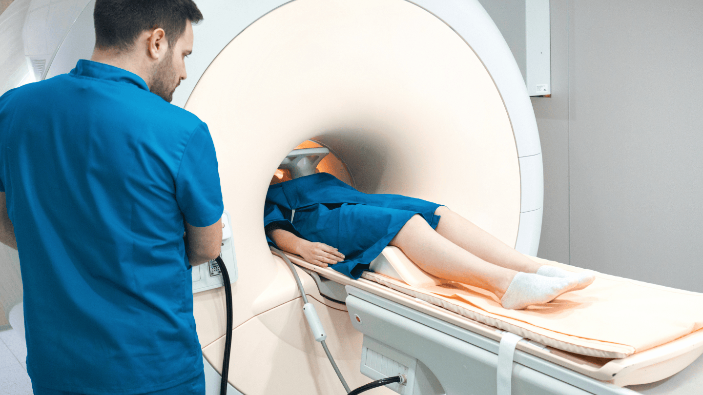
An MRI scan is a type of imaging test that uses powerful magnets and radio waves instead of radiation to generate detailed images of your body's internal structures. Unlike X-rays, which are particularly effective at showing bones, MRI scans excel at imaging soft tissues, such as brains, muscles, blood vessels, ligaments and organs.
You can think of an MRI scan as taking many thin "slices" through your body, similar to slicing a loaf of bread, with each "slice" showing a different layer. The MRI machine can create 2D cross-sectional images and 3D images that can be viewed from multiple angles. Your healthcare provider can use the images to diagnose and treat various medical conditions.
The traditional MRI scanner is a large, cylindrical machine that acts like a giant magnet. This can make some people feel uncomfortable or claustrophobic. To address this, several variations of MRI are available, such as:
Open MRI, which has openings on three sides to reduce the enclosed feeling
Short bore MRI, which is more wider and shorter than traditional scanners
How does it work?
To create an image of your internal structures, the MRI machine first generates a strong magnetic field. It causes the hydrogen atoms in the water and fat throughout your body to align with this magnetic field because of their natural magnetic properties.
The MRI machine then sends a pulse of radio waves into your body. For a brief moment, this pulse causes the hydrogen atoms to become misaligned. When the radio pulse is turned off, the hydrogen atoms gradually return to their original position.
As they realign, they emit faint radio signals. Sensors inside the MRI scanner detect these signals from different parts of your body. A computer then processes and analyses these signals to construct detailed cross-sectional images of your internal structure.
The scan usually lasts between 30 and 60 minutes but can sometimes last up to 90 minutes. During the scan, you can expect the following:
You will be asked to lie down on a table that slides into the MRI scanner.
When the scan starts, the MRI machine will make loud tapping or thumping sounds. You'll be given earplugs or headphones to wear before the procedure to protect your hearing.
During the scan, it's important to stay still to ensure a clear image. You may occasionally be asked to hold your breath as well.
If there are issues, you can use the intercom to contact the MRI technologist in another room.
When is MRI best used?
MRI scans excel at imaging soft tissues and provide clear images of these structures, often better than X-rays. Doctors commonly use them to diagnose and monitor a broad range of medical conditions throughout your body. These conditions include:
Brain and nervous system conditions:
Brain tumours and other abnormal growths
Stroke and other blood vessel abnormalities in the brain
Multiple sclerosis and other degenerative diseases
Spinal cord injuries and compression
Herniated discs and other spinal problems
Musculoskeletal injuries and conditions:
Ligament and tendon tears, particularly in the knee, shoulder, and ankle
Cartilage damage and meniscus tears
Arthritis and other joint inflammatory conditions
Muscle injuries
Bone infections
Heart and blood vessel assessment:
Heart disease and other heart conditions
Blocked or narrowed blood vessels
Congenital heart defects and structural abnormalities
Internal organ examination:
Liver diseases, including cirrhosis and liver cancer
Kidney abnormalities and tumours
Pancreatic conditions and inflammation
Breast abnormalities (often used alongside mammogram)
Prostate conditions
Pelvic conditions, including uterine and ovarian issues
Cancer detection and monitoring:
Detecting tumours throughout the body
Staging cancer to determine extent and spread
Monitoring treatment response during chemotherapy or radiation therapy
Cancer treatment follow-up examinations
Planning surgical treatment
Since the MRI uses no ionising radiation, it's considered very safe for repeated examinations when necessary. This makes it the preferred imaging choice when frequent monitoring is necessary.
To find out more about MRI and X-ray scans, schedule a consultation with Thomson Medical. Our specialist can provide you more information about this procedure and decide which scan is best for you.
What is the difference between X-rays and MRIs?
Both X-rays and MRIs are important diagnostic imaging techniques employed by your doctor, but they work in different ways and serve different functions. Understanding these differences helps to ensure that you receive the most suitable imaging for your medical needs.
The following summary outlines the differences between the two tests:
| MRI | X-ray | |
|---|---|---|
| How it works | Uses strong magnets and radio waves to create detailed images | X-ray radiation is used to create an image |
| Radiation exposure | No radiation exposure | Small amounts of exposure to radiation, which is generally safe but can accumulate with repeated scans |
| When is the best time to use it? | Assessing soft tissues, such as:
| It is the most effective method for generating detailed images of bones. Also commonly used to assess:
|
| Common medical uses | Detecting conditions such as:
| Detecting conditions such as:
|
| Duration | The duration depends on the area examined, ranging from 30 to 90 minutes | The duration depends on the area examined, ranging from a few minutes to 10 minutes |
| Cost in Singapore | The cost depends on the type of MRI being performed, but it is generally pricier than an X-ray. For example, an MRI scan of the lumbar spine can cost up to SGD 1,000, whereas a full-body MRI can cost as much as SGD 2,500. | The cost depends on the type of X-ray being performed, but it's generally cheaper than an MRI scan. For example, a dental X-ray can cost as little as SGD 40, while a mammogram can cost up to SGD 200. |
| Safety |
|
|
| Availability | Due to its size, an MRI machine is only available in a large medical centre or hospital | Due to their relatively small size, they are commonly available at emergency care centres, polyclinics, or dental clinics |
| Disadvantages |
|
|
Which one should you choose?
Neither MRI nor X-ray imaging is better than the other. Each has its advantages and limitations, making them suitable for different medical situations. The most appropriate imaging method for you will depend on your specific medical needs, as well as the area of your body requiring examination.
Your healthcare provider will recommend the most appropriate imaging method for you, ensuring you receive the most accurate diagnosis possible.
To discuss which test suits your condition, schedule a consultation with Thomson Medical. Our specialist can help provide you with more information about this procedure, its benefits, and any potential risks.
FAQ
What will an MRI show that an X-ray will not?
An MRI provides detailed images of soft tissues, such as muscles, ligaments, tendons, the brain, and internal organs, structures that X-rays cannot visualise well. This makes MRI essential for diagnosing conditions like torn ligaments, brain tumours, and spinal cord injuries.
What can an MRI not detect?
MRIs are less effective for detecting bone fractures compared to X-rays. They may also struggle to identify small calcium deposits or air-filled structures like lungs.
Why would a doctor order an MRI or an X-ray?
Your doctor may order an X-ray to assess bone injuries, joint dislocations, lung conditions, or dental issues. While an MRI scan is usually chosen to evaluate soft tissue damage, neurological conditions, or complex joint and spinal injuries.
Does an MRI show broken bones?
While MRIs can detect bone fractures, X-rays are usually the first choice for diagnosing most bone injuries due to their efficiency and accuracy in visualising bone structures.
Why would a doctor not order an MRI?
Your doctor may avoid ordering an MRI scan if an X-ray can provide a clear diagnosis, if you have metal implants or certain medical devices, or if the cost and availability of an MRI scan make it impractical for a minor condition.
Which is more expensive, an MRI or an X-ray?
MRI scans are generally much pricier than X-rays. In Singapore, an MRI can cost from SGD 1000 to over 2,500, while X-rays typically range from SGD 40 to 200, depending on the type of scan.
The information provided is intended for general guidance only and should not be considered medical advice. To get personalised recommendations based on your medical conditions, schedule an appointment with Thomson Medical.
For more information, contact us:
Thomson Medical Concierge
- 8.30am - 5.30pm
- WhatsApp: 9147 2051
Need help finding the right specialist or booking for a group?
Our Medical Concierge is here to help you. Simply fill in our form, and we'll check and connect you with the right specialist promptly.
Notice:
The range of services may vary between Thomson clinic locations. Please contact your preferred branch directly to enquire about the current availability.
Get In Touch