
Many people might think a single spine MRI scan covers everything—but it actually doesn’t. Radiologists typically scan one spinal region at a time for a clearer image and more accurate results.
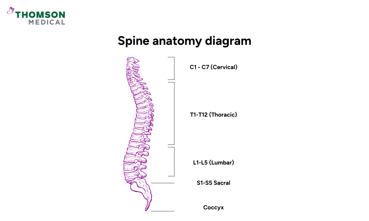
Based on the spiral anatomy, these regions are divided into three main types, which are lumbar (lower back), cervical (neck), and thoracic (mid-back). This guide will guide you through the differences between these spinal regions, so you’ll know what to expect before stepping into the scanner.
What is a spine MRI scan?
A spine MRI (magnetic resonance imaging) is a non-invasive imaging test that uses a strong magnetic field and radio waves—without radiation—to produce high-resolution images of the spine, spinal cord, discs, and surrounding soft tissues.
It helps doctors to diagnose conditions such as herniated discs, nerve compression, spinal stenosis, and other spinal abnormalities. Unlike X-rays or CT scans, an MRI does not use ionising radiation, which makes it a safer option for repeated imaging.
Types of spine MRI scans
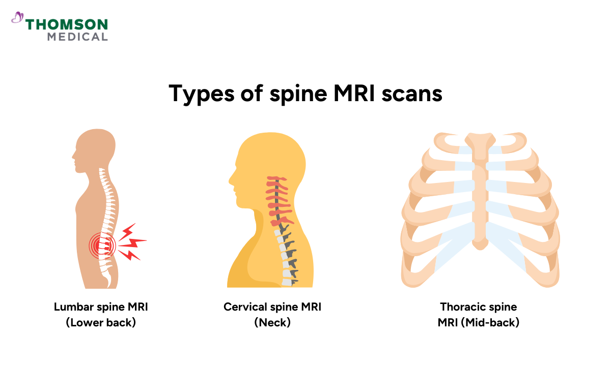
Spine MRIs are typically divided into three main types based on the anatomical region being examined. Each scan provides detailed pictures of specific spinal structures to help identify the underlying cause of symptoms such as pain, numbness, weakness, or limited mobility.
Lumbar spine MRI (lower back)
The lumbar MRI focuses on the lower portion of the spine, typically from L1 to L5 vertebrae. This scan is most often recommended for patients experiencing chronic lower back pain, leg weakness, or symptoms of sciatica. It can detect:
Herniated or bulging discs pressing on nerves
Lumbar spinal stenosis (narrowing of the spinal canal)
Degenerative disc disease
Nerve root compression (radiculopathy)
Inflammatory or infectious conditions
Vertebral fractures or alignment issues
It is particularly useful for assessing conditions that affect the sciatic nerve or cause lower body discomfort, especially in individuals with sedentary lifestyles or a history of heavy lifting.
Cervical spine MRI (neck)
A cervical MRI captures detailed images of the neck region, covering vertebrae C1 through C7. This type of scan is ideal for investigating neck pain, headaches, or neurological symptoms radiating from the shoulders, arms, or hands. It helps diagnose:
Cervical disc herniation or degeneration
Spinal cord compression
Cervical spondylosis or arthritis
Pinched nerves (cervical radiculopathy)
Whiplash injuries or trauma
Tumours or cysts near the cervical spine
It’s especially important in cases where symptoms include tingling, numbness, or muscle weakness in the upper limbs.
Thoracic spine MRI (mid-back)
The thoracic spine MRI evaluates the mid-back, including vertebrae T1 to T12. Although less commonly scanned than the cervical or lumbar regions, the thoracic spine is crucial when investigating more complex or serious conditions. It can reveal:
Compression fractures (often from osteoporosis or trauma)
Spinal cord lesions or inflammation
Thoracic disc disease (though rare)
Tumours in or near the spinal cord
Multiple sclerosis plaques or demyelinating disorders
Infections such as discitis or osteomyelitis
This scan is often done when a patient has upper back pain, nerve-related symptoms, or signs of illness that doctors can't easily explain.
If you have been advised to undergo a spine MRI scan or would like to learn more about this medical imaging and your options, you can request an appointment with us to schedule a consultation.
When should you have a spine MRI?
A spinal MRI may be recommended if you’re experiencing persistent back, neck, or nerve-related symptoms that do not improve with rest or initial treatment.
This imaging test helps doctors identify the underlying cause and guide appropriate care. You may need a spine MRI if you have:
Chronic back or neck pain lasting more than a few weeks
Numbness, tingling, or weakness in the arms or legs
Symptoms of sciatica or nerve compression
Injury from a fall, accident, or trauma
Loss of bladder or bowel control (may signal spinal cord compression)
Suspected spinal infection, inflammation, or tumour
Monitoring of conditions like multiple sclerosis or spinal arthritis
Always consult your doctor to determine whether a spine MRI is necessary based on your specific symptoms and medical history.
A consultation with a healthcare provider can help determine whether a spine MRI is needed to better understand the cause and guide your treatment plan. Request an appointment with Thomson Medical to speak with a qualified healthcare provider and receive further evaluation based on your medical conditions.
Why should you consider a spine MRI?
Spine MRI scans are commonly performed to help healthcare professionals accurately diagnose a wide range of spinal conditions and neurological symptoms.
It provides detailed images of the spinal cord, vertebrae, discs, and surrounding tissues—making it a valuable tool when X-rays or CT scans do not provide enough information. Therefore, doctors may tell you to do a spine MRI to investigate:
Herniated or bulging discs:
An MRI for lumbar herniated disc can detect disc material pressing against spinal nerves, a common cause of persistent back or neck pain.
Pinched nerves (sciatica or radiculopathy):
Compression of spinal nerve roots may result in pain, numbness, tingling, or weakness radiating to the arms or legs.
Spinal cord compression:
This condition causes compression of the spinal cord, which can lead to walking difficulties, coordination problems, or loss of bladder and bowel control.
Spinal injuries:
MRI can evaluate damage caused by trauma, falls, or accidents—including ligament tears, fractures, or disc injury not visible on X-ray.
Degenerative spine diseases and arthritis:
MRI shows signs of wear and tear such as disc degeneration, bone spurs, or facet joint arthritis, which may contribute to chronic pain or stiffness.
Spinal tumours or infections:
The scan helps detect abnormal growths, inflammation, or spinal abscesses that may indicate infection, cancer, or other underlying conditions.
Multiple sclerosis (MS):
Spine MRI is often used alongside brain MRI to identify lesions or plaques on the spinal cord that are characteristic of multiple sclerosis.
If you are experiencing ongoing spine-related symptoms, request an appointment with Thomson Medical. Our qualified specialists can help to determine whether an MRI is appropriate for your condition.
How to prepare for a spine MRI?

Proper preparation helps ensure your spine MRI is safe, accurate, and stress-free. Here’s what you should keep in mind before your scan:
Wear loose, metal-free clothing:
Choose clothing without zippers, buttons, or metal fastenings. Jewellery, watches, and piercings should also be removed prior to the scan.
Inform your doctor about any implants or medical devices:
Let your healthcare provider know if you have metal implants, pacemakers, or joint replacements, as some metal objects may interfere with scan images.
Follow dietary instructions if provided:
Most spine MRIs do not require fasting, so you can usually eat and drink as normal. However, always follow any specific guidelines given by your doctor or imaging centre.
Discuss claustrophobia or anxiety in advance:
If you feel anxious in confined spaces, speak with your doctor beforehand. You may be offered a mild sedative or referred for an open MRI if appropriate.
Notify your doctor if you are pregnant or breastfeeding:
While MRI scans are generally considered safe during pregnancy, your doctor will assess the benefits and potential risks before proceeding.
What to expect during a spine MRI scan?
During the scan, you’ll lie flat on a motorised table that slowly slides into the MRI machine. To capture more detailed images, a specialised coil may be positioned around your neck or back. It’s important to remain as still as possible to ensure image clarity.
You might hear loud knocking or thumping sounds while being scanned—this is normal. Earplugs or headphones will be provided to reduce noise and help you feel more comfortable. Typically, the scan process itself took around 30 to 60 minutes to complete.
In some cases, a contrast dye may be injected into a vein to highlight certain structures more clearly. While the scan itself is painless, some individuals may feel slightly uncomfortable due to the confined space or sounds. If you have any concerns, let your care team know beforehand.
Risks with spine MRI scans
Spine MRIs are considered very safe, especially since they do not involve radiation. However, there are a few precautions and mild risks to be aware of:
Metal implants or devices:
Patients with pacemakers, metal implants, or some hearing assist devices (cochlear implants) must inform their doctor, as some devices can interfere with MRI safety or image quality.
Contrast dye reactions (if used):
In some cases, a contrast agent (gadolinium) may be injected to enhance image clarity. Side effects are rare but may include mild nausea, headache, metallic taste, or temporary itching.
Claustrophobia or discomfort:
Some individuals may feel anxious in the enclosed MRI scanner. If you're concerned about confined spaces, discuss this with your doctor beforehand—alternatives or medication may be offered to ease discomfort.
The cost of a spine MRI scan in Singapore
The cost of a spine MRI scan in Singapore varies depending on several factors, such as the area of the spine being scanned (for example cervical, thoracic, or lumbar), use of contrast dye, consultation fee with the specialist, and whether the scan is done at a public or private facility.
At subsidised rates in public hospitals, a spine MRI scan typically costs between SGD 500 and SGD 1,200, depending on the complexity and body region scanned. Prices may be higher for non-subsidised patients or those without a referral from a public clinic.
In private hospitals or imaging centres, the cost can range from SGD 1,000 to over 2,500, especially if contrast dye is used or a detailed consultation with a spine specialist is included.
Factors that may affect the final cost include:
Specific region of the spine scanned (e.g., lumbar vs whole spine)
Use of contrast-enhanced imaging
Inclusion of specialist consultation or follow-up
Public vs private healthcare facility
The information provided above is intended for general reference only. For detailed fee information and payment options, please consult your healthcare provider directly. Request an appointment with our specialists at Thomson Medical for a detailed price breakdown and a personalised care plan.
FAQ
What does a spine MRI show?
A spine MRI captures detailed images of your spinal cord, vertebrae, discs, nerves, and surrounding tissues. It helps doctors diagnose:
- Herniated or bulging discs, which are discs pressing on nerves causing pain or stiffness
- Pinched nerves, which are linked to numbness, weakness, or sciatica
- Spinal cord issues, like inflammation, infection, or multiple sclerosis
- Degenerative diseases, such as arthritis or spinal stenosis
- Injuries, such as from falling, trauma, or accidents
- Tumours or cysts, which are anabnormal growths along the spine
What organs are visible on a lumbar MRI?
A lumbar MRI mainly focuses on the lower spine but may also show nearby structures, including:
- Spine, discs, and nerves
- Muscles and ligaments
- Blood vessels
- Pelvic organs, such as parts of the kidneys, bladder, uterus, or prostate
How long does a spine MRI take?
A spine MRI typically takes 30 to 60 minutes, depending on the scan area and whether contrast dye is needed. You’ll need to lie still throughout the procedure, but you can breathe normally. If contrast is used, the scan may take slightly longer.
Do you lie on your back for a spine MRI?
Yes, for most spine MRI scans, you will lie on your back on a padded table that slides into the MRI machine. Depending on the scan type (lumbar, cervical, or thoracic), a special coil may be placed around the area being scanned to improve image quality. In some cases, alternative positions may be used based on medical needs, but lying on your back is the most common.
Is a spine MRI painful?
No, a spine MRI is not painful. You simply lie still while the machine captures images. Some people may feel discomfort from lying flat or being in a confined space, but breaks can be provided if needed. If you’re claustrophobic, your doctor may offer a mild sedative to help you stay calm during the scan.
Can a spinal MRI show nerve damage?
A spine MRI can reveal nerve compression, irritation, or inflammation, often caused by:
- Herniated or bulging discs
- Spinal stenosis (narrow spinal canal)
- Inflammation or swelling around nerves
- Tumours or cysts pressing on nerve tissue
However, it does not measure nerve function. If functional nerve damage is suspected, your doctor may order additional tests like EMG (electromyography) to assess how well the nerves are working.
The information provided is intended for general guidance only and should not be considered medical advice. For personalised recommendations and tailored advice, please consult a specialist at Thomson Medical. Request an appointment with Thomson Medical today.
For more information, contact us:
Thomson Medical Concierge
- 8.30am - 5.30pm
- WhatsApp: 9147 2051
Need help finding the right specialist or booking for a group?
Our Medical Concierge is here to help you. Simply fill in our form, and we'll check and connect you with the right specialist promptly.
Notice:
The range of services may vary between Thomson clinic locations. Please contact your preferred branch directly to enquire about the current availability.
Get In Touch