
You've been waiting to see your baby's face, and the images from regular ultrasounds leave you wanting more. Some expectant parents choose 3D ultrasound scans to get that first real glimpse of their baby's features – but you're probably wondering if it's worth it, when's the best time to go, and whether it's safe.
The decision isn't just about getting a clearer picture; it's about understanding what these scans can and can't do, how they fit into your prenatal care, and whether this optional experience is the right choice for your pregnancy journey.
What is a 3D ultrasound scan?
A 3D ultrasound gives three-dimensional pictures of your unborn little one inside the womb. While traditional 2D ultrasounds show flat, black-and-white images, 3D ultrasounds capture images from multiple angles and combine them to show depth, giving you a more lifelike picture of your baby's features.
How is it different from 2D and 4D scans?
The 3D ultrasound uses the same soundwave technology as a standard ultrasound scan, but the images are processed by a computer to form a three-dimensional view. The difference is in how the images are assembled together – rather than showing one flat slice at a time, a computer assembles many images together to form a 3D view.
2D ultrasound:
A 2D ultrasound provides flat, cross-sectional images of the baby and is most commonly used in routine pregnancy care for checking foetal growth, heartbeat, and development.
3D ultrasound:
A 3D ultrasound, in contrast, creates lifelike, three-dimensional still images in a single frozen frame. This allow parents and doctors to see the baby’s face, limbs, and external features in more detail.
4D ultrasound:
The fourth dimension – time – distinguishes 4D ultrasound from its 3D counterpart. A 4D ultrasound goes one step further by adding real-time movement, essentially showing a live video of the baby moving in the womb.
When is a 3D ultrasound performed during pregnancy?
The ideal time window to go for 3D ultrasounds is between 26 and 32 weeks of pregnancy (second or third trimester). This timing gives the clearest images because your baby has developed enough facial features and body definition to show up well, while there's still enough amniotic fluid and space in your uterus for good imaging.
Why does timing matters?
If you go too early for scan, your baby's features may not be developed enough to see clearly. If you wait too long to get your scan done, your baby gets larger and has less room to move, making it harder to capture clear images – especially the face of your baby.
What affects image quality?
Even at an optimal time, several factors can affect imaging:
Baby's position:
If your baby is facing your back or has their hands covering their face, it's harder to get good facial images
Placenta location:
If the placenta is in front of your baby's face, it can block the view
Amniotic fluid levels:
Too much or too little fluid can affect image clarity
Baby's movement:
Sometimes babies cooperate, sometimes they don't
Your sonographer may ask you to move around, take a walk, or come back another day if your baby isn't in a good position for imaging.
What can you see in a 3D ultrasound?
A 3D ultrasound provides a three-dimensional view of your baby's external features with remarkable detail that standard 2D scans cannot capture. Instead of the flat, cross-sectional images you see in traditional ultrasounds, 3D technology creates lifelike images that show depth and contours.
So you will see the following features in the 3D scan:
Facial features:
Nose shape, lips, profile, and sometimes even facial expressions
Hands and feet:
Individual fingers and toes, hand positions
Body structure:
Overall body proportions and positioning
The three-dimensional view helps visualise external anatomy more clearly than standard ultrasound, making it easier to see how features are forming and developing.
Why is a 3D foetal ultrasound done?
3D ultrasounds serve both medical and personal purposes, though they are optional rather than routine prenatal care.
Medical reasons
Your doctor may recommend a 3D ultrasound scan if they need detailed visualisation of suspected abnormalities identified during standard 2D scans:
Cleft lip or palate
Spinal cord defects (spina bifida)
Limb abnormalities or malformations
Facial structural issues
Abdominal wall defects
The three-dimensional view helps your pregnancy doctor confirm or rule out these external structural conditions more definitively than 2D imaging alone.
Personal reasons
Many parents choose 3D ultrasounds simply to see their baby's face and features more clearly. This can be a meaningful bonding experience, especially for partners who want to feel more connected to the pregnancy. Seeing your baby's realistic features, such as their nose, lips, and expressions, creates memorable moments that families often treasure.
However, while 3D ultrasounds provides detailed images and can assist with specific diagnoses, they don't replace routine 2D scans. Standard 2D ultrasound is essential for monitoring internal organ development, measuring growth, assessing blood flow, and evaluating overall foetal health throughout pregnancy.
Interested in seeing your baby in 3D? Our obstetricians and gynaecologists can help you determine the optimal timing for your scan and answer any questions about the process. You can contact Thomson Medical to arrange a consultation and receive guidance tailored to your needs.
Our O&G specialists
Loading...
What to expect during the appointment?
A 3D ultrasound works just like your regular pregnancy scans. Here is how it normally goes:
You'll lie back on an examination table, and then the sonographer applies gel to your belly to help sound waves travel clearly.
The sonographer will then move an ultrasound probe (hand-held device used during the scan) over your abdomen to capture images.
The ultrasound system then processes the sound waves into three-dimensional images of your baby.
You can watch the images appear on screen in real-time as the scan happens.
If your baby isn't cooperating (facing away, hands covering face), you may need to move around, take a short walk, or reschedule for clearer images.
The procedure is safe, painless, and non-invasive. Most scans take about 20 to 45 minutes, and you’ll usually be able to watch your baby’s images in real time on the screen. Many clinics also provide printed or digital keepsake pictures, allowing you to take home a memory of the experience.
Is a 3D foetal ultrasound safe?
Yes, a 3D foetal ultrasound is considered safe when it is performed by qualified professionals. It uses sound waves to create images and does not expose the baby or mother to radiation.
Unlike X-rays or CT scans, ultrasounds are non-invasive and radiation-free. They have been used in pregnancy for decades with no evidence of harming the baby. However, medical professionals recommend that an ultrasound scanning should only be performed when there is a medical need, rather than repeating scans without reason.
Limitations of a 3D scan
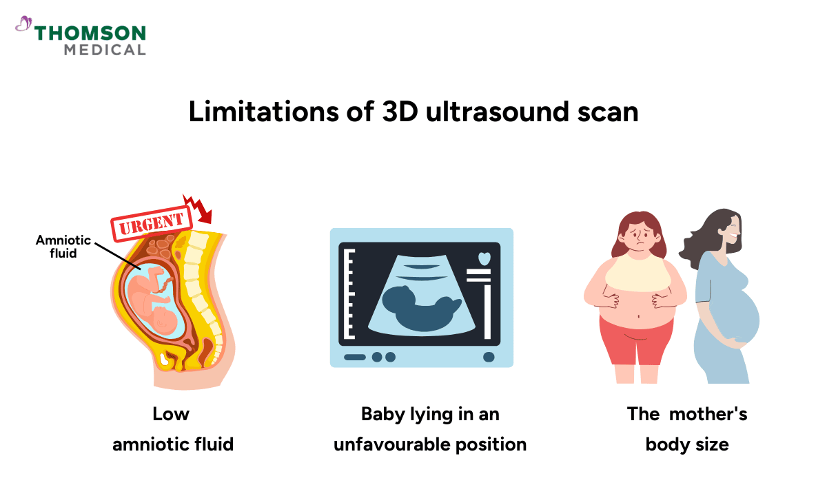
While a 3D ultrasound can provide clear, lifelike images of your baby, there are a few natural limitations to keep in mind:
The quality of images depends on the position of the baby, the amount of the amniotic fluid, the stage of pregnancy, and even the mother's body type.
If the baby is turned to face the uterus wall or has arms or legs in front of their face, the images may not be very clear.
While external features are well represented, 3D scans are not used to examine internal organs or look at overall growth, which can be assessed better by a standard 2D ultrasound.
3D ultrasounds are often recommended as a complement and are not employed as a regular diagnostic tool in prenatal care.
FAQ
Is 3D foetal ultrasound necessary during pregnancy?
No, 3D ultrasounds are not medically necessary for most pregnancies. Standard 2D ultrasounds provide all the essential information doctors need to monitor your baby's growth, heartbeat, and organ development and detect most abnormalities. Your doctor may recommend a 3D scan if they need a closer look at specific features like facial structures or limbs or to evaluate a suspected concern more thoroughly.
Can I get a 3D ultrasound during my first trimester?
3D ultrasounds aren't recommended in the first trimester because your baby is too small for 3D imaging. Standard obstetric ultrasound in the first trimester can effectively confirms pregnancy location, detects the heartbeat, and dates your pregnancy. The optimal time for 3D ultrasound is between 26 and 32 weeks, when your baby's facial features are well-developed.
Will a 3D ultrasound detect abnormalities?
3D ultrasound imaging can provide additional views of certain physical features, but they're not primary diagnostic tools. Your regular 2D ultrasounds remain the standard for medical monitoring. Your pregnancy doctors may use 3D imaging alongside other tests when they need more detailed visualisation of specific areas.
What happens if the images aren’t clear?
If images aren't clear, your sonographer may ask you to change positions, gently massage your abdomen, or wait a few minutes for the baby to move. Sometimes walking around or drinking water encourages movement. If the images remain unclear, the clinic may schedule another appointment on a different day.
It is important to remember that even if the 3D images are not perfect, standard 2D ultrasounds can still provide all the necessary medical information about your baby’s health.
Is 3D ultrasound the same as my prenatal screening tests?
No, 3D ultrasounds are separate from routine prenatal screening tests like your anomaly scan or blood work. Standard 2D ultrasounds and prenatal screening tests provide all necessary medical information about your baby's health, while 3D scans are primarily for bonding and keepsake ultrasound photos.
How much does a 3D ultrasound cost?
3D ultrasound costs vary depending on your location and the package you choose, as they're typically not covered by insurance for non-medical purposes. Contact your preferred ultrasound centre directly for current pricing information. Many facilities offer different packages based on session length and number of images or videos included.
The information provided is intended for general guidance only and should not be considered medical advice. For personalised recommendations and tailored advice, please consult a specialist at Thomson Medical. Request an appointment with Thomson Medical today.
For more information, contact us:
Thomson Specialists (Women's Health)
Thomson Women's Clinic (TWC)
- Novena:
6592 6686 (Call), 8611 8986 (WA) - Bukit Batok:
6569 0668 (Call), 8686 3525 (WA) - Choa Chu Kang:
6893 1227 (Call), 8282 1796 (WA) Jurong:
6262 8588 (Call), 6262 8588 (WA)- Katong (female doctor):
6970 2272 (Call), 8611 9020 (WA) - Punggol:
6243 6843 (Call), 8811 0328 (WA) - Sembawang: 6753 5228
- Sengkang: 6388 8125
- Serangoon (female doctor): 6382 3313
- Tampines: 6857 6266
- Tiong Bahru: 6276 1525