
Cervical cancer is the fourth most common cancer among women worldwide.
The good news is that it can often be prevented with regular screening. Finding it early significantly improves the chances of successful treatment, which is why early detection is so important—not just for your health, but for your peace of mind.
However, many women still experience fear and uncertainty when it comes to a possible diagnosis. Learning how tools like ultrasound and other tests help detect cervical cancer can give you confidence and encourage you to take charge of your health.
The role of ultrasound in detecting cervical cancer
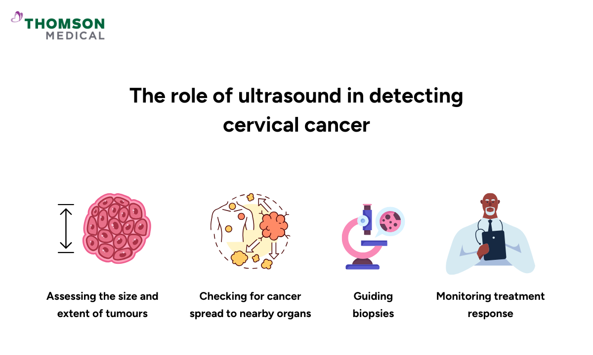
Ultrasound is a non-invasive imaging test that uses high-frequency sound waves to create pictures of internal organs. While it is a valuable tool in many areas of gynaecology, ultrasound alone cannot reliably detect cervical cancer in its earliest stages. Instead, it plays a supportive role in cervical cancer diagnosis and management, including:
Assessing the size and extent of tumours:
Ultrasound can measure the dimensions of visible cervical tumours, giving doctors insight into how much of the cervix is affected.
Checking for cancer spread to nearby organs:
Transvaginal ultrasound can help determine if cancer has extended into surrounding tissues such as the bladder or rectum.
Guiding biopsies:
Ultrasound imaging can assist doctors in precisely targeting suspicious areas when taking tissue samples.
Monitoring treatment response:
During and after treatment, ultrasound may be used to track tumour size and evaluate how well therapies are working.
Because ultrasound has limitations in detecting early-stage cervical cancer, doctors typically rely on other diagnostic tools such as ThinPrep Pap test, Pap smears, HPV testing, MRI, or CT scans for accurate diagnosis and staging.
How ultrasound works in cervical cancer
Ultrasound, also called sonography, uses sound waves to create images of internal organs. For cervical evaluation, two main techniques are used:
The probe is placed on the lower abdomen. This method gives a general view of the pelvis but is less detailed for small cervical changes.
A small probe is inserted into the vagina, providing a much closer and clearer image of the cervix, uterus, and surrounding tissues.
This broader scan combines transabdominal and/or transvaginal approaches to assess the entire female reproductive system, including the cervix, uterus, ovaries, and fallopian tubes. It is commonly used when symptoms such as pelvic pain, abnormal bleeding, or suspected tumours need further investigation.
Because the cervix is located deep in the pelvis, transvaginal ultrasound is the preferred method when doctors need to investigate cervical abnormalities.
Why ultrasound alone cannot diagnose cervical cancer
While ultrasound can show suspicious findings, it cannot confirm whether these changes are cancerous. Many non-cancerous conditions – such as cervical polyps, fibroids, or infections – may appear abnormal on ultrasound.
To confirm the diagnosis, a biopsy (tissue sample) is always required.
Clinical role of ultrasound
Early suspicion:
If a patient presents with symptoms like abnormal bleeding or pelvic pain, ultrasound is often one of the first diagnostic imaging tools used.
Staging tool:
In confirmed cases, ultrasound helps determine the size and spread of the tumour, which is crucial for staging cervical cancer.
Treatment guidance:
Ultrasound assists in planning surgery or radiotherapy and can also monitor tumour response during or after treatment.
Advantages and limitations of ultrasound in detecting cervical cancer
While it offers many benefits, it also has limitations that affect its role in screening and diagnosis.
Advantages of ultrasound
Safe and non-invasive:
Ultrasound uses sound waves rather than radiation, making it safe for repeated use, even in sensitive cases such as pregnancy.
It does not require incisions, injections of contrast dye, or exposure to harmful rays.
Widely available and cost-effective:
Ultrasound machines are accessible in most hospitals and clinics, including in low-resource settings where advanced imaging like MRI or PET scans may not be available.
It is usually less expensive than other imaging modalities, making it more affordable for patients.
Real-time imaging:
Ultrasound provides immediate images, allowing doctors to see movements, blood flow, and tissue changes as they occur.
This is particularly useful for guiding procedures such as biopsies, where precision is important.
Useful in staging and monitoring:
Once cervical cancer is suspected or diagnosed, ultrasound helps measure tumour size and assess spread to surrounding tissues or lymph nodes.
It can also be used during follow-up visits to evaluate whether a tumour is responding to treatment or whether it has returned (recurrence).
Comfortable for most patients:
While transvaginal ultrasound may cause slight discomfort, the procedure is generally well tolerated and does not require anaesthesia.
Limitations of ultrasound
Not suitable for early detection:
Ultrasound cannot reliably detect very early cervical cancer or precancerous changes in cervical cells.
Operator-dependent accuracy:
The quality of the images and their interpretation depends heavily on the skill and experience of the sonographer or doctor performing the scan.
Inexperienced operators may miss subtle signs of disease.
Limited detail compared to other scans:
Although useful, ultrasound does not provide the same level of detail as MRI, which is the gold standard for staging cervical cancer.
Small tumours or microscopic disease spread may not be visible on ultrasound.
Cannot confirm diagnosis:
Ultrasound can show abnormal masses or tissue changes but cannot confirm whether they are cancerous.
A biopsy is always needed for a definitive diagnosis.
Restricted by patient factors:
Image quality can be affected by obesity, scarring, or the presence of bowel gas, making it more difficult to obtain clear pictures.
Therefore, ultrasound can be considered as a safe, affordable, and valuable tool in the assessment and management of cervical cancer, particularly for staging and treatment monitoring. However, it is not effective as a stand-alone screening test and should always be used in combination with cervical screening programmes and biopsy for accurate diagnosis.
Other imaging methods for cervical cancer
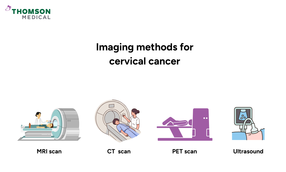
While ultrasound has a role in monitoring cervical cancer in later stages, it is not the most reliable method for initial diagnosis. Several other imaging techniques are considered more accurate and effective in detecting, staging, and monitoring cervical cancer:
MRI (Magnetic resonance imaging)
MRI scans produce highly detailed images of soft tissues.
Considered the gold standard for assessing tumour size and spread within the cervix.
Plays a key role in treatment planning.
Offers greater accuracy than ultrasound for evaluating cervical tumours.
CT (Computed tomography) scan
CT scan helps determine whether cervical cancer has spread to nearby lymph nodes or distant organs.
Commonly used for staging and treatment planning.
Tracks treatment response over time.
Provides more detail than ultrasound when examining surrounding organs.
PET (Positron emission tomography) scan
PET scan is often combined with CT (PET-CT) for comprehensive imaging.
Detects areas where cancer may have spread beyond the cervix.
Useful for monitoring how effective treatment is.
Can highlight cancer activity that might not be visible on MRI, CT, or ultrasound alone.
MRI is generally the most effective imaging technique for detecting and staging cervical cancer, while CT and PET scans provide vital information about cancer spread and treatment response. Ultrasound, although limited, may still be used in specific situations to complement these advanced methods.
How do doctors diagnose cervical cancer?
Diagnosing cervical cancer usually involves several steps, beginning with routine screening and moving to more detailed investigations if abnormalities are found.
Initial screening tests
Pap smear: Collects cells from the cervix for laboratory analysis to detect abnormal or precancerous changes.
HPV test: Identifies high-risk strains of the human papillomavirus (HPV), which are strongly linked to the development of cervical cancer. Persistent HPV infection increases risk.
Diagnostic procedures
Colposcopy: Colposcopy is a closer examination of the cervix using a special microscope to identify abnormal areas.
Biopsy: Removal of tissue samples to confirm whether cancer cells are present. Types include:
Punch biopsy: Takes small samples from the cervix.
Cone biopsy: Removes a larger, cone-shaped piece of cervical tissue.
LEEP (Loop Electrosurgical Excision Procedure): Uses a thin wire loop to remove abnormal tissue for testing.
A Pap smear or HPV test may detect early changes, but only a biopsy can confirm cervical cancer. Imaging tests are then used to understand its spread and guide treatment planning.
Doctors who can help diagnose cervical cancer
Loading...
Early detection and the right imaging approach can make a life-changing difference. If you’re concerned about cervical cancer or have been advised to undergo imaging, request an appointment with Thomson Medical and we will help you determine which test is most appropriate for your situation.
FAQ
What is the biggest indicator of cervical cancer?
The most common and important warning sign of cervical cancer is abnormal vaginal bleeding. This can include:
Bleeding between periods
Bleeding after sexual intercourse
Heavier or longer menstrual bleeding than usual
Bleeding after menopause
Other possible symptoms may include unusual vaginal discharge, pelvic pain, or pain during sex.
Can doctors see cervical cancer on ultrasound?
Yes, doctors can sometimes see cervical cancer on ultrasound, especially if the tumour is large enough to alter the shape or structure of the cervix. Ultrasound can show abnormal growths, changes in tissue, or unusual blood flow patterns. However, it cannot always detect very early cervical cancer, and it cannot confirm whether an abnormality is cancerous. A biopsy is always required for a definite diagnosis.
Can the cervix be seen on ultrasound?
Yes, the cervix can be seen on ultrasound. A transabdominal scan may show it, but a transvaginal ultrasound gives a much clearer and more detailed view, making it the preferred method for examining the cervix.
Is a transvaginal ultrasound painful?
A transvaginal ultrasound may cause mild discomfort, but it should not be painful. The procedure is usually quick and well tolerated.
What is the most accurate test for detecting cervical cancer?
The Pap smear and HPV test are the most effective for early detection. If results are abnormal, colposcopy and biopsy are performed for confirmation.
How accurate is ultrasound compared to a Pap smear?
Ultrasound is helpful for evaluating tumour size, spread, and staging once cancer is suspected, but Pap smears and HPV testing are much more accurate for early detection.
When should you start screening for cervical cancer, and how often?
Most guidelines recommend starting cervical cancer screening at age 25. From ages 25 to 64, women and people with a cervix should be screened every 3 to 5 years, depending on the type of test offered in their country (Pap smear, HPV testing, or both).
Ages 25–49: usually every 3 years
Ages 50–64: usually every 5 years
Over 65: screening is often not needed if recent results have been normal, unless there are new symptoms or risk factors.
Screening may start earlier or be done more frequently for those at higher risk, such as people with HIV, a weakened immune system, or a history of abnormal cervical results. Therefore, regular screening is the best way to detect cervical changes early, before they develop into cancer.
The information provided is intended for general guidance only and should not be considered medical advice. For personalised recommendations and tailored advice based on your unique situations, please consult a specialist at Thomson Medical. Request an appointment with Thomson Medical today.
For more information, contact us:
Thomson Specialists (Women's Health)
Thomson Women's Clinic (TWC)
- Novena:
6592 6686 (Call), 8611 8986 (WA) - Bukit Batok:
6569 0668 (Call), 8686 3525 (WA) - Choa Chu Kang:
6893 1227 (Call), 8282 1796 (WA) Jurong:
6262 8588 (Call), 6262 8588 (WA)- Katong (female doctor):
6970 2272 (Call), 8611 9020 (WA) - Punggol:
6243 6843 (Call), 8811 0328 (WA) - Sembawang: 6753 5228
- Sengkang: 6388 8125
- Serangoon (female doctor): 6382 3313
- Tampines: 6857 6266
- Tiong Bahru: 6276 1525